Light Microscope Collection (#8)

Exploring the Intricacies of Life: A Glimpse through the Light Microscope Unveiling the Wonders of Cerebellum Tissue

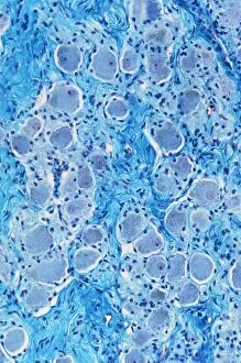
Background imageLight Microscope Collection: Nerve ganglion, light micrograph C016 / 0532

Nerve ganglion, light micrograph C016 / 0532
Nerve ganglion. Light micrograph of a section through a nerve ganglion of the peripheral nervous system, showing clusters of nerve cell bodies

Background imageLight Microscope Collection: Myelinated nerve fibres, light micrograph C016 / 0531

Myelinated nerve fibres, light micrograph C016 / 0531
Myelinated nerve fibres. Light micrograph of a section through a spinal sensory nerve containing many closely packed axons exhibiting a wave-type appearance

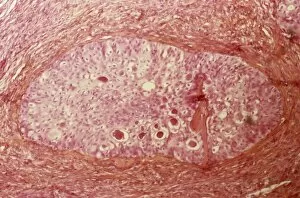
Background imageLight Microscope Collection: Brenner tumour of the ovary C015 / 7137

Brenner tumour of the ovary C015 / 7137
Brenner tumour of the ovary. Light micrograph of a section through an ovary showing a Brenner tumour (centre). This rare benign (non-cancerous)

Background imageLight Microscope Collection: Colon, light micrograph C016 / 0516

Colon, light micrograph C016 / 0516
Colon. Light micrograph of a section through the mucosa (inner epithelial lining) of the colon (large bowel), showing the typical arrangement of invaginations of columnar epithelial cells forming

Background imageLight Microscope Collection: Langerhans cell histiocytosis C015 / 7135

Langerhans cell histiocytosis C015 / 7135
Langerhans cell histiocytosis. Light micrograph of a section through a lymph node showing Langerhans cell histiocytosis (LCH), also known as granulomatosis

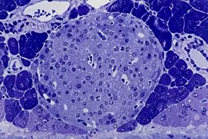
Background imageLight Microscope Collection: Islet of Langerhans, light micrograph C016 / 0514

Islet of Langerhans, light micrograph C016 / 0514
Islet of Langerhans. Light micrograph of a section through an islet of Langerhans in a pancreas. Scattered throughout the pancreas, the islets are endocrine cells that secrete a range of hormones

Background imageLight Microscope Collection: Blood cells, light micrograph C015 / 7130

Blood cells, light micrograph C015 / 7130
Blood cells. Light micrograph of red blood cells (erythrocytes, red) and white blood cells (leucocytes, nuclei stained pink)

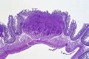
Background imageLight Microscope Collection: Bowel cancer, light micrograph C016 / 0511

Bowel cancer, light micrograph C016 / 0511
Bowel cancer. Light micrograph of a section through a carcinoid tumour in the small bowel. The tumour has invaded the submucosa of the bowel

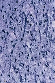
Background imageLight Microscope Collection: Brain glial cells, light micrograph C016 / 0523

Brain glial cells, light micrograph C016 / 0523
Brain glial cells. Light micrograph of a section through brain tissue, showing numerous glial cells (dark). Glial cells, or neuroglia, are non-neural cells of the central nervous system (CNS)

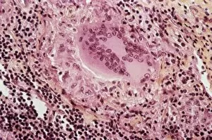
Background imageLight Microscope Collection: Sarcoidosis, light micrograph C015 / 7134

Sarcoidosis, light micrograph C015 / 7134
Sarcoidosis. Light micrograph of a section through a lymph node affected by sarcoidosis. Also known as Boecks disease, this is a chronic inflammation of the lymph nodes or organs

Background imageLight Microscope Collection: Lung silicosis, light micrograph C015 / 7129

Lung silicosis, light micrograph C015 / 7129
Lung silicosis. Light micrograph of a section through a lung with silicosis, a type of pneumoconiosis caused by exposure to silica dusts. The lung tissue has become fibrous (centre)

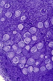
Background imageLight Microscope Collection: Ovarian primordial follicles, micrograph C016 / 0518

Ovarian primordial follicles, micrograph C016 / 0518
Ovarian primordial follicles. Light micrograph of a section through the cortex of a neonatal ovary, showing clusters of primordial follicles each containing a single oocyte (egg cell)
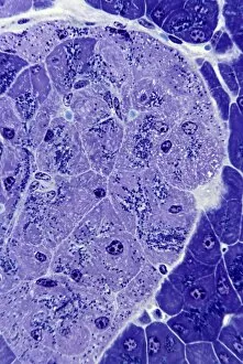
Background imageLight Microscope Collection: Islet of Langerhans, light micrograph C016 / 0513

Islet of Langerhans, light micrograph C016 / 0513
Islet of Langerhans. Light micrograph of a section through an islet of Langerhans in a pancreas. Scattered throughout the pancreas, the islets are endocrine cells that secrete a range of hormones

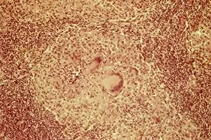
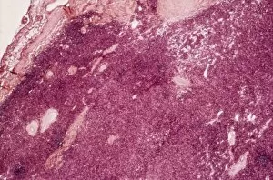
Background imageLight Microscope Collection: Thymus gland cancer, light micrograph C015 / 7128

Thymus gland cancer, light micrograph C015 / 7128
Thymus cancer. Light micrograph of a section through a thymus gland with thymoma cancer. The thymus is a gland in the upper chest that plays an important role in the immune system

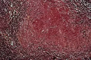
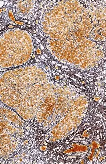
Background imageLight Microscope Collection: Cirrhosis of liver, light micrograph C016 / 0529

Cirrhosis of liver, light micrograph C016 / 0529
Cirrhosis of liver. Light micrograph of a section through liver tissue, damaged by cirrhosis. Cirrhosis occurs as a result of a range of factors causing damage to liver function

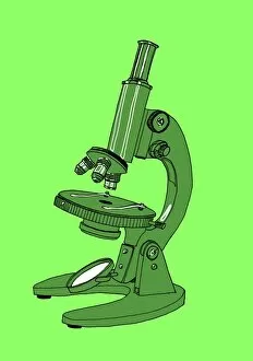
Background imageLight Microscope Collection: Light microscope, illustration C018 / 0737

Light microscope, illustration C018 / 0737
Light microscope. Illustration of a light microscope with three lenses and a mirror for reflecting light towards the object being viewed

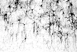
Background imageLight Microscope Collection: Cerebral cortex nerve cells C018 / 0062

Cerebral cortex nerve cells C018 / 0062
Cerebral cortex nerve cells. Light micrograph of a section through neurones (nerve cells, black) in the cerebral cortex of a human brain

Background imageLight Microscope Collection: Light microscope, illustration C018 / 0738

Light microscope, illustration C018 / 0738
Light microscope. Illustration of a light microscope with three lenses and a mirror for reflecting light towards the object being viewed

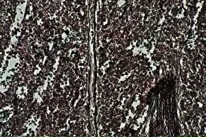
Background imageLight Microscope Collection: Spleen, light micrograph C014 / 0858

Spleen, light micrograph C014 / 0858
Spleen. Light micrograph of a section through the red pulp of a spleen. Situated on the left side of the upper abdomen, behind the stomach, the spleen is a large lymphoid organ

Background imageLight Microscope Collection: Cerebrum, light micrograph C014 / 1399

Cerebrum, light micrograph C014 / 1399
Cerebrum. Light micrograph of a section through tissue from the cerebrum, showing neurons (nerve cells, purple). The cerebrum is the largest part of the brain

Background imageLight Microscope Collection: Cerebrum, light micrograph C014 / 1400

Cerebrum, light micrograph C014 / 1400
Cerebrum. Light micrograph of a section through tissue from the cerebrum, showing neurons (nerve cells, purple). The cerebrum is the largest part of the brain

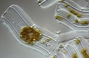
Background imageLight Microscope Collection: Diatoms, light micrograph C014 / 4675

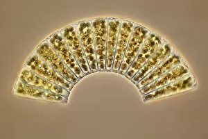
Diatoms, light micrograph C014 / 4675
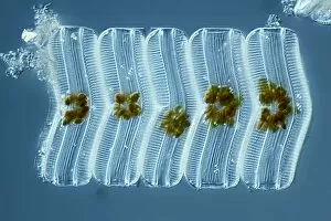
Diatoms. Differential interference contrast micrograph of a group of Meridion circulare diatoms. Diatoms are a group of photosynthetic, single-celled algae containing about 10, 000 species

Background imageLight Microscope Collection: Poppy seeds, light micrograph C014 / 4658

Poppy seeds, light micrograph C014 / 4658
Poppy (Papaver sp.) seeds, light micrograph. Magnification: x34, when printed 10 centimetres wide

Background imageLight Microscope Collection: Cat fluke, light micrograph C014 / 4858

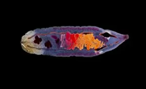
Cat fluke, light micrograph C014 / 4858
Cat fluke (Opisthorchis felineus), light micrograph. This parasitic trematode (flatworm) infests the liver of members of the cat family

Background imageLight Microscope Collection: Grass seed, light micrograph C014 / 4727

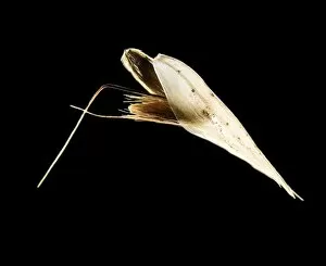
Grass seed, light micrograph C014 / 4727
Anopheles mosquito. Coloured scanning electron micrograph (SEM) of an Anopheles stephensi mosquito, the vector for the malaria parasite in Asia

Background imageLight Microscope Collection: Diatom, light micrograph C014 / 4677

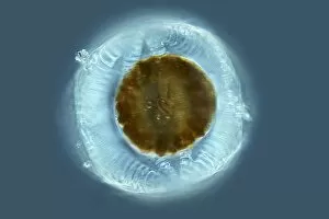
Diatom, light micrograph C014 / 4677
Diatom. Differential interference contrast micrograph of a Campylodiscus sp. diatom. Diatoms are a group of photosynthetic, single-celled algae containing about 10, 000 species

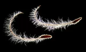
Background imageLight Microscope Collection: Greater burdock seeds, light micrograph C014 / 4728

Greater burdock seeds, light micrograph C014 / 4728
Greater burdock (Arctium lappa) seeds. Light micrograph of two fruits (achenes) from a greater burdock plant. Magnification: x7.5 when printed 10 cm wide

Background imageLight Microscope Collection: Water beetle, light micrograph C014 / 4881

Water beetle, light micrograph C014 / 4881
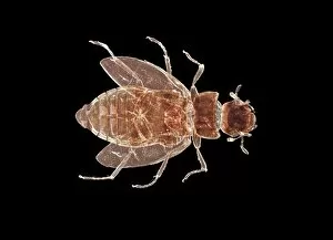
Water beetle. Light micrograph showing the anatomy of a small water beetle (order Coleoptera)

Background imageLight Microscope Collection: Green hydra, light micrograph C014 / 4680

Green hydra, light micrograph C014 / 4680
Green hydra. Darkfield illuminated light micrograph of the tentacled head of a green hydra (Hydra viridis). Hydra are small simple predatory fresh-water animals, belonging to the phylum Cnidaria

Background imageLight Microscope Collection: Green algae, light micrograph C014 / 4670

Green algae, light micrograph C014 / 4670
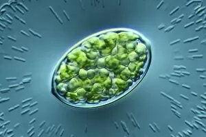
Green algae. Differential interference contrast micrograph of a single green alga, showing the chloroplast (green) inside. Algae use the chloroplasts to produce food through photosynthesis

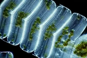
Background imageLight Microscope Collection: Diatoms, light micrograph C014 / 4671

Diatoms, light micrograph C014 / 4671
Diatoms. Differential interference contrast micrograph of a group of Achnantes longipes diatoms. Diatoms are a group of photosynthetic, single-celled algae containing about 10, 000 species

Background imageLight Microscope Collection: Green hydra, light micrograph C014 / 4681

Green hydra, light micrograph C014 / 4681
Green hydra. Darkfield illuminated light micrograph of a green hydra (Hydra viridis), showing its tentacled head. Hydra are small simple predatory fresh-water animals

Background imageLight Microscope Collection: Rove beetle, light micrograph C014 / 4873

Rove beetle, light micrograph C014 / 4873
Rove beetle. Dark-field illuminated light micrograph of a Stenus sp. rove beetle

Background imageLight Microscope Collection: Tulipa sp. flower, light micrograph C014 / 4654

Tulipa sp. flower, light micrograph C014 / 4654
Tulip (Tulipa sp.) flower. Light micrograph of a tulip flower, showing the male anthers (dark) and female stigma (centre). Magnification: x17 when printed 10 centimetres wide

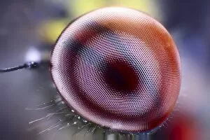
Background imageLight Microscope Collection: Damselfly eye, light micrograph C014 / 4664

Damselfly eye, light micrograph C014 / 4664
Damselfly eye. Light micrograph showing one of the compound eyes of a damselfly (order Odonata). Each compound eye is made up of numerous simple eyes called ommatidia

Background imageLight Microscope Collection: 19th Century bullseye condenser

19th Century bullseye condenser and stand. Bullseye condensers contain a lens (upper right) that is used to focus and control the light illuminating a subject under a microscope. Made in 1870

Background imageLight Microscope Collection: White dryas seeds, light micrograph C014 / 4733

White dryas seeds, light micrograph C014 / 4733
White dryas (Dryas octopetala) seeds. Light micrograph of two fruits (achenes) from a white dryas plant. Magnification: x7.5 when printed 10 cm wide

Background imageLight Microscope Collection: Methyl sulphonal crystals, micrograph

Methyl sulphonal crystals, micrograph
Methyl sulphonal crystals, polarised light micrograph. Magnification: x114 when printed 10 centimetres wide

Background imageLight Microscope Collection: Beelouse, light micrograph C014 / 4841

Beelouse, light micrograph C014 / 4841
Beelouse (Braula coeca), dark-field illuminated light micrograph. This wingless fly is a parasite of the honeybee (Apis mellifera)

Background imageLight Microscope Collection: Diatom, light micrograph C014 / 4669

Diatom, light micrograph C014 / 4669
Diatom. Differential interference contrast micrograph of an Achnantes longipes diatom. Diatoms are a group of photosynthetic, single-celled algae containing about 10, 000 species

Background imageLight Microscope Collection: Fish louse C014 / 4837

Fish louse C014 / 4837
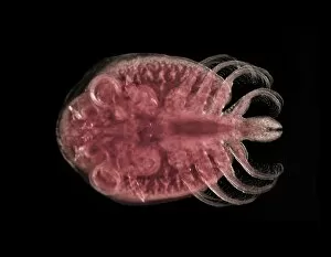
Fish louse. Dark-field illuminated light micrograph of a fish louse (Argulus sp.). This freshwater crustacean is a serious pest in commercial fish farms and fisheries

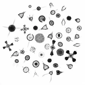
Background imageLight Microscope Collection: Radiolaria, light micrograph C014 / 4672

Radiolaria, light micrograph C014 / 4672
Radiolaria. Collection of light micrographs showing various different species of radiolaria. Radiolaria (or radiolarians) are amoeboid protozoa that produce intricate mineral skeletons

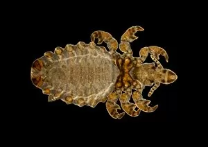
Background imageLight Microscope Collection: Parasitic louse, light micrograph C014 / 4850

Parasitic louse, light micrograph C014 / 4850
Parasitic louse (Haematopinus asini), light micrograph. This louse commonly infects horses. It causes irritation that can cause damage by the horse rubbing itself raw

Background imageLight Microscope Collection: Paraneplocephala tapeworm, micrograph C014 / 4859

Paraneplocephala tapeworm, micrograph C014 / 4859
Paraneplocephala tapeworm. Light micrograph of a Paraneplocephala sp. tapeworm. This short parasitic tapeworm (order Cestoidea) is found in horses and other herbivores

Background imageLight Microscope Collection: Plant seedling, light micrograph C014 / 4741

Plant seedling, light micrograph C014 / 4741
Plant seedling, light micrograph. Magnification: x5 when printed 10 cm wide

Background imageLight Microscope Collection: Diatoms, light micrograph C014 / 4668

Diatoms, light micrograph C014 / 4668
Diatoms. Differential interference contrast micrograph of a group of Achnantes longipes diatoms. Diatoms are a group of photosynthetic, single-celled algae containing about 10, 000 species
Background imageLight Microscope Collection: Diatoms, light micrograph C014 / 4674

Diatoms, light micrograph C014 / 4674
Diatoms. Differential interference contrast micrograph of a group of Achnantes longipes diatoms. Diatoms are a group of photosynthetic, single-celled algae containing about 10, 000 species

Background imageLight Microscope Collection: Green hydra, light micrograph C014 / 4678

Green hydra, light micrograph C014 / 4678
Green hydra. Light micrograph of the tentacled head of a green hydra (Hydra viridis). Hydra are small simple predatory fresh-water animals, belonging to the phylum Cnidaria


For sale as Licensed Images

Choose your image, Select your licence and Download the media

Exploring the Intricacies of Life: A Glimpse through the Light Microscope Unveiling the Wonders of Cerebellum Tissue: Witness the intricate network of cells in cerebellum tissue as seen through a captivating light micrograph. Nerve and Glial Cells Revealed: Dive into the mesmerizing world of nerve and glial cells, captured in stunning detail by a powerful light microscope. Unraveling Chemical Structures: Behold the ethereal beauty of copper and magnesium sulphate crystals, magnified under a light micrograph to expose their delicate arrangements. The Hidden Beauty Within Caffeine Crystals: Marvel at the crystalline structures of caffeine, brought to life through an enchanting light micrograph that unveils its hidden allure. Journey into Hippocampus Brain Tissue: Embark on an extraordinary expedition deep within hippocampus brain tissue, where mysteries unfold with every glance through our remarkable light microscope lens. Illuminating Glial Cells' Secrets: Delve into the enigmatic realm of glial cells as they reveal their intricate patterns and connections in a mesmerizing confocal light micrograph. HeLa Cells Exposed - A Window to Immortality? Explore HeLa cells like never before with our cutting-edge technology, providing unprecedented insight into these immortalized wonders under brightfield illumination (C017 / 8299). Peering Into Purkinje Nerve Cells in Cerebellum: Discover the elegance and complexity displayed by Purkinje nerve cells residing within cerebellar tissue when observed using a high-resolution light microscope. Dicotyledon Plant Stem's Inner Workings Revealed. Get lost amidst nature's marvels as we uncover the intricacies present within dicotyledon plant stems using our advanced techniques for capturing breathtakingly detailed images under brightfield illumination. Unveiling the Secrets of Kidney Tubules.