Light Microscope Collection (page 5)

Exploring the Intricacies of Life: A Glimpse through the Light Microscope Unveiling the Wonders of Cerebellum Tissue

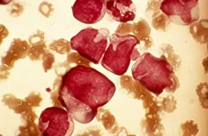
Background imageLight Microscope Collection: Acute myeloid leukaemia, micrograph

Acute myeloid leukaemia, micrograph
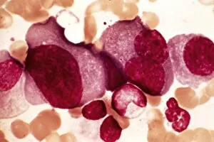
Acute myeloid leukaemia. Light micrograph of blood cells from bone marrow in a case of acute myeloid leukaemia. These cells include a monocytoid myeloblast stem cell

Background imageLight Microscope Collection: Elliptocytosis, light micrograph

Elliptocytosis, light micrograph
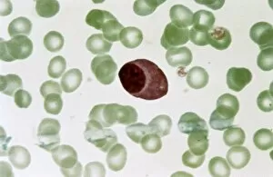
Elliptocytosis. Light micrograph of red blood cells in a case of elliptocytosis. Red blood cells (erythrocytes) carry oxygen and carbon dioxide to and from body tissues
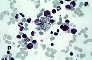
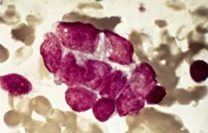
Background imageLight Microscope Collection: Cystine in bone marrow, light micrograph

Cystine in bone marrow, light micrograph
Cystine in bone marrow. Light micrograph of crystals of cystine among blood cells in a sample of bone marrow. Cystine is an amino acid that can form crystals in urine

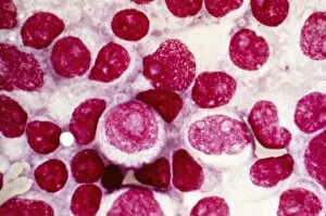
Background imageLight Microscope Collection: Bone marrow blood cells, light micrograph

Bone marrow blood cells, light micrograph

Background imageLight Microscope Collection: Ciliate protozoan ingesting algae

Ciliate protozoan ingesting algae. Darkfield illuminated light micrograph of a ciliate protozoan ingesting green algae (green)

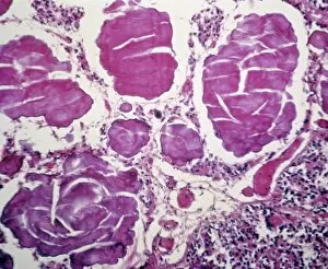
Background imageLight Microscope Collection: Parkinsons disease, light micrograph

Parkinsons disease, light micrograph
Parkinsons disease. Light micrograph of a section through the sabstantia nigra of the brain of a patient with Parkinsons disease showing a neuron (nerve cell)

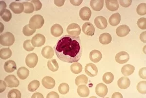
Background imageLight Microscope Collection: Monocyte blood cell, light micrograph

Monocyte blood cell, light micrograph. Monocytes are a type of white blood cell, part of the bodys immune system. Monocytes are the largest white blood cell

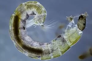
Background imageLight Microscope Collection: Midge larva, light micrograph

Midge larva, light micrograph
Midge larva. Differential interference contrast (DIC) micrograph of the larva of a non-biting midge (family Chironomidae). Midge refers to many kinds of very small two-winged flies found world-wide

Background imageLight Microscope Collection: Chebarkul meteorite, light micrograph C015 / 2863

Chebarkul meteorite, light micrograph C015 / 2863
Chebarkul meteorite fragment seen through a microscope in polarised light. This research is being carried out at the Vernadsky Institute, Moscow, Russia

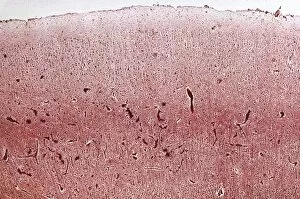
Background imageLight Microscope Collection: Dementia, light micrograph

Dementia, light micrograph
Dementia. Light micrograph of a section through the brain of a patient with dementia. Dementia is mainly a disease of old age

Background imageLight Microscope Collection: Desmid, light micrograph

Desmid, light micrograph
Desmid. Darkfield illuminated light micrograph of a desmid green alga. Desmids are a common group of freshwater single-celled algae that have intricate cell walls

Background imageLight Microscope Collection: Pernicious anaemia, light micrograph

Pernicious anaemia, light micrograph
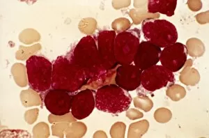
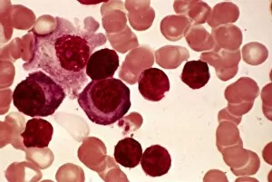
Pernicious anaemia. Light micrograph of megaloblast blood cells from bone marrow in a case of pernicious anaemia, also known as Biermers anaemia

Background imageLight Microscope Collection: Blood cell cancer, light micrograph

Blood cell cancer, light micrograph
Blood cell cancer. Light micrograph of blood cells from a lymphatic ganglion in a case of a blood cancer of a mixed cell type

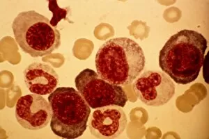
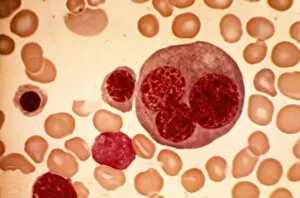
Background imageLight Microscope Collection: Erythroblast blood cell, light micrograph

Erythroblast blood cell, light micrograph
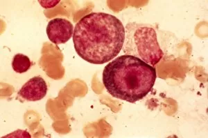
Erythroblast blood cell. Light micrograph of cells from a sample of bone marrow, including a polychromatic erythroblast (upper left). This one has multiple cell nuclei (dark red, four in total)

Background imageLight Microscope Collection: Infectious mononucleosis, micrograph

Infectious mononucleosis, micrograph
Infectious mononucleosis. Light micrograph of blood cells obtained from an inflamed lymphatic ganglion in a case of infectious mononucleosis (glandular fever)

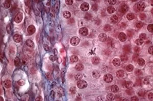
Background imageLight Microscope Collection: Corpus luteum, light micrograph

Corpus luteum, light micrograph
Corpus luteum. Light micrograph of a section through the corpus luteum of an ovary. The corpus luteum is the tissue formed from a mature ovarian follicle after the follicle has released its oocyte

Background imageLight Microscope Collection: Haematococcus alga, light micrograph

Haematococcus alga, light micrograph
Haematococcus alga. Differential interference contrast (DIC) micrograph of a Haematococcus sp. green alga. Magnification: x670, when printed 10 centimetres wide

Background imageLight Microscope Collection: Oocyte before ovulation, light micrograph

Oocyte before ovulation, light micrograph
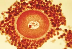
Oocyte before ovulation. Light micrograph of a section through tissue from an ovary, showing an oocyte two days before ovulation

Background imageLight Microscope Collection: Euastrum desmid, light micrograph

Euastrum desmid, light micrograph
Euastrum desmid. Darkfield illuminated light micrograph of a Euastrum oblongum desmid green alga. Desmids are a common group of freshwater single-celled algae that have intricate cell walls

Background imageLight Microscope Collection: Lymphoblastic sarcoma, light micrograph

Lymphoblastic sarcoma, light micrograph
Lymphoblastic sarcoma. Light micrograph of blood cells from bone marrow in a case of lymphoblastic sarcoma. These small lymphoblasts (dark red) characteristically have conspicuous nucleoli

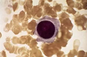
Background imageLight Microscope Collection: Plasmocyte blood cell, light micrograph

Plasmocyte blood cell, light micrograph. Plasmocytes, also called plasma cells, are derived from stimulated B lymphocytes (white blood cells)

Background imageLight Microscope Collection: Xanthidium desmids, light micrograph

Xanthidium desmids, light micrograph
Xanthidium desmids. Polarised light micrograph of a Xanthidium sp. desmid green algae. Desmids are a common group of freshwater single-celled algae that have intricate cell walls

Background imageLight Microscope Collection: Multiple myeloma, light micrograph

Multiple myeloma, light micrograph
Multiple myeloma. Light micrograph of dystrophy (abnormality) of plasmocyte blood cells from bone marrow in a case of multiple myeloma, also known as Kahlers disease or plasma cell myeloma

Background imageLight Microscope Collection: Desmids, light micrograph

Desmids, light micrograph
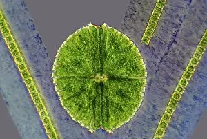
Desmids. Phase contrast light micrograph of Micrasterias sp. and Hyalotheca sp. desmid green algae. Desmids are a common group of freshwater single-celled algae that have intricate cell walls

Background imageLight Microscope Collection: Cushings syndrome, light micrograph

Cushings syndrome, light micrograph
Cushings syndrome. Light micrograph of a section through muscle affected by Cushings syndrome, a hormonal condition caused by overproduction of adrenal gland hormones

Background imageLight Microscope Collection: Lymphoplasmacytic lymphoma, micrograph

Lymphoplasmacytic lymphoma, micrograph
Lymphoplasmacytic lymphoma. Light micrograph of a mixture of small lymphocytes and plasma cells from bone marrow in a case of lymphoplasmacytic lymphoma, also known as Waldenstroms macroglobulinemia

Background imageLight Microscope Collection: Ovarian follicle tissue, light micrograph

Ovarian follicle tissue, light micrograph
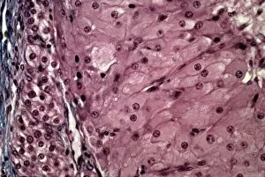
Ovarian follicle tissue. Light micrograph of a section through tissue from an ovarian follicle, showing a boundary between two layers

Background imageLight Microscope Collection: Hairy cell leukaemia, light micrograph

Hairy cell leukaemia, light micrograph
Hairy cell leukaemia. Light micrograph of blood cells in a case of hairy cell leukaemia. This sample, treated with May-Grunwald-Giemsa (MGG) stain

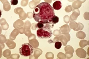
Background imageLight Microscope Collection: Mast cells, light micrograph

Mast cells, light micrograph
Mast cells. Light micrograph of several mast cells (pink rounded objects). These leucocytes (white blood cells) contain the chemical mediators histamine, serotonin and heparin

Background imageLight Microscope Collection: Chebarkul meteorite fragment C015 / 2862

Chebarkul meteorite fragment C015 / 2862
Chebarkul meteorite fragment seen through a microscope. This research is being carried out at the Vernadsky Institute, Moscow, Russia

Background imageLight Microscope Collection: Microalgae, light micrograph

Microalgae, light micrograph
Microalgae. Differential interference contrast (DIC) micrograph of Botryococcus braunii (round), and other species of green alga. Magnification: x278, when printed 10 centimetres wide

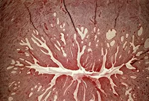
Background imageLight Microscope Collection: Cervix, light micrograph

Cervix, light micrograph
Cervix. Light micrograph of a section through the mucosa of the cervix, the neck of the uterus. This is the endocervix, which borders the uterus. It is lined with columnar epithelial cells

Background imageLight Microscope Collection: Acute erythroid leukaemia, micrograph

Acute erythroid leukaemia, micrograph
Acute erythroid leukaemia. Light micrograph of blood cells from bone marrow in a case of acute erythroid leukaemia. The cells include dystrophic (degenerated) erythroblasts (nuclei stained dark red)

Background imageLight Microscope Collection: Closterium desmid, light micrograph

Closterium desmid, light micrograph
Closterium desmid. Differential interference contrast (DIC) micrograph of a Closterium sp. desmid green alga. Desmids are a common group of freshwater single-celled algae that have intricate cell

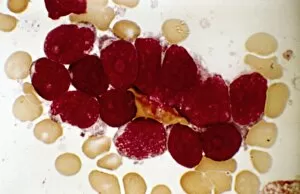
Background imageLight Microscope Collection: Acute lymphoblastic leukaemia, micrograph

Acute lymphoblastic leukaemia, micrograph
Acute lymphoblastic leukaemia. Light micrograph of blood cells from bone marrow in a case of acute lymphoblastic leukaemia. Leukaemia is a cancer where certain blood cells form in excess

Background imageLight Microscope Collection: Malignant histiocytosis, light micrograph

Malignant histiocytosis, light micrograph
Malignant histiocytosis. Light micrograph of blood cells in a case of malignant histiocytosis. Hystiocytosis is an excess number of histiocytes

Background imageLight Microscope Collection: Haematococcus algae, light micrograph

Haematococcus algae, light micrograph
Haematococcus algae. Differential interference contrast (DIC) micrograph of a colony of Haematococcus sp. green alga. Magnification: x220, when printed 10 centimetres wide

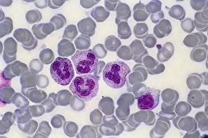
Background imageLight Microscope Collection: Blood cells, light micrograph

Blood cells, light micrograph
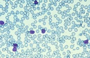
Blood cells. Light micrograph of red blood cells (erythrocytes, light blue) and white blood cells (leucocytes, nuclei stained purple)

Background imageLight Microscope Collection: Chediak-Higashi syndrome, micrograph

Chediak-Higashi syndrome, micrograph
Chediak-Higashi syndrome. Light micrograph of blood cells in a case of Chediak-Higashi syndrome. The cells include neutrophil white blood cells with abnormal granules

Background imageLight Microscope Collection: Metastatic breast cancer, micrograph

Metastatic breast cancer, micrograph
Metastatic breast cancer. Light micrograph of blood cells in a case of metastatic (secondary) medullary breast cancer. Medullary breast cancer is a rare form of breast cancer that is diagnosed by

Background imageLight Microscope Collection: Ciliate protozoan, light micrograph

Ciliate protozoan, light micrograph
Ciliate protozoan. Darkfield illuminated light micrograph of a ciliate protozoan, showing green algae (green) that it has ingested

Background imageLight Microscope Collection: Metastatic lung cancer, micrograph

Metastatic lung cancer, micrograph
Metastatic lung cancer. Light micrograph of blood cells in a case of metastatic (secondary) medullary lung cancer. Medullary cancer (carcinoma)

Background imageLight Microscope Collection: Multiple sclerosis, light micrograph

Multiple sclerosis, light micrograph
Multiple sclerosis. Light micrograph of a section through the brain showing a large area of demyelinated nerve fibres (light pink, top) due to multiple sclerosis

Background imageLight Microscope Collection: Pineal gland, light micrograph

Pineal gland, light micrograph. The pineal gland is a small endocrine gland located near to the centre of the brain, between the two hemispheres


For sale as Licensed Images

Choose your image, Select your licence and Download the media

Exploring the Intricacies of Life: A Glimpse through the Light Microscope Unveiling the Wonders of Cerebellum Tissue: Witness the intricate network of cells in cerebellum tissue as seen through a captivating light micrograph. Nerve and Glial Cells Revealed: Dive into the mesmerizing world of nerve and glial cells, captured in stunning detail by a powerful light microscope. Unraveling Chemical Structures: Behold the ethereal beauty of copper and magnesium sulphate crystals, magnified under a light micrograph to expose their delicate arrangements. The Hidden Beauty Within Caffeine Crystals: Marvel at the crystalline structures of caffeine, brought to life through an enchanting light micrograph that unveils its hidden allure. Journey into Hippocampus Brain Tissue: Embark on an extraordinary expedition deep within hippocampus brain tissue, where mysteries unfold with every glance through our remarkable light microscope lens. Illuminating Glial Cells' Secrets: Delve into the enigmatic realm of glial cells as they reveal their intricate patterns and connections in a mesmerizing confocal light micrograph. HeLa Cells Exposed - A Window to Immortality? Explore HeLa cells like never before with our cutting-edge technology, providing unprecedented insight into these immortalized wonders under brightfield illumination (C017 / 8299). Peering Into Purkinje Nerve Cells in Cerebellum: Discover the elegance and complexity displayed by Purkinje nerve cells residing within cerebellar tissue when observed using a high-resolution light microscope. Dicotyledon Plant Stem's Inner Workings Revealed. Get lost amidst nature's marvels as we uncover the intricacies present within dicotyledon plant stems using our advanced techniques for capturing breathtakingly detailed images under brightfield illumination. Unveiling the Secrets of Kidney Tubules.