
Light Microscope Collection (page 7)
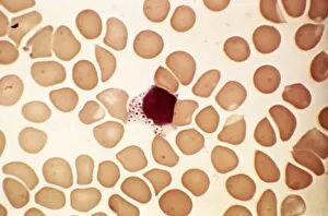
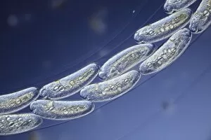
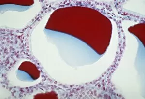
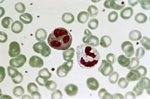
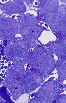
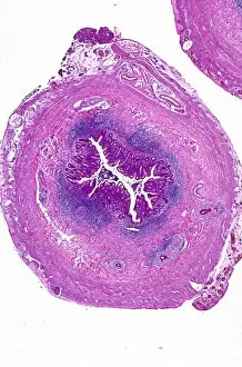
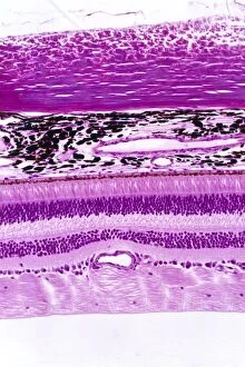
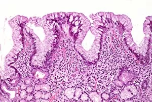
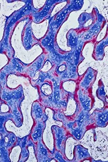
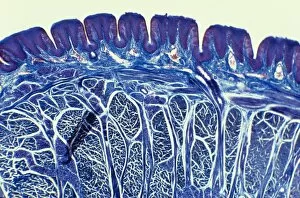
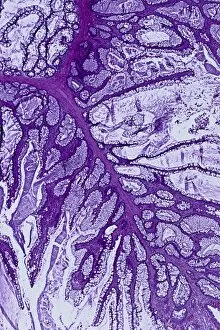
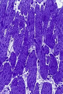
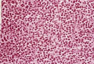
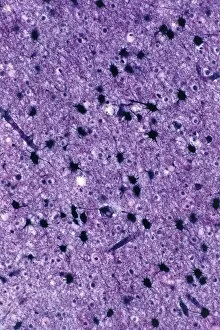
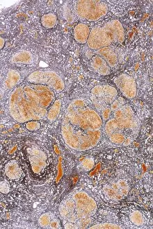
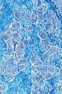
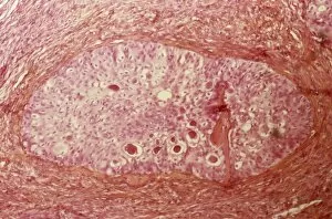
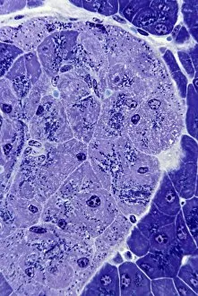
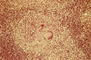
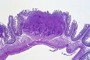
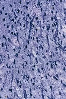
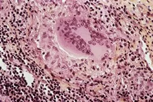
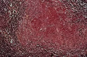
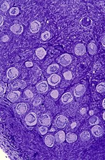
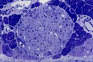
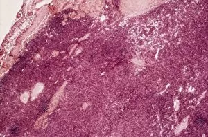
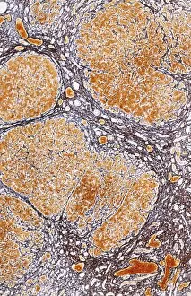
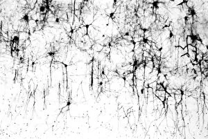
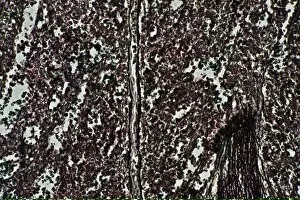
Exploring the Intricacies of Life: A Glimpse through the Light Microscope Unveiling the Wonders of Cerebellum Tissue


For sale as Licensed Images
Choose your image, Select your licence and Download the media
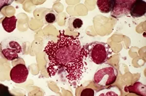
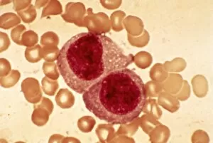
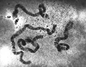
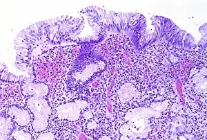
Exploring the Intricacies of Life: A Glimpse through the Light Microscope Unveiling the Wonders of Cerebellum Tissue: Witness the intricate network of cells in cerebellum tissue as seen through a captivating light micrograph. Nerve and Glial Cells Revealed: Dive into the mesmerizing world of nerve and glial cells, captured in stunning detail by a powerful light microscope. Unraveling Chemical Structures: Behold the ethereal beauty of copper and magnesium sulphate crystals, magnified under a light micrograph to expose their delicate arrangements. The Hidden Beauty Within Caffeine Crystals: Marvel at the crystalline structures of caffeine, brought to life through an enchanting light micrograph that unveils its hidden allure. Journey into Hippocampus Brain Tissue: Embark on an extraordinary expedition deep within hippocampus brain tissue, where mysteries unfold with every glance through our remarkable light microscope lens. Illuminating Glial Cells' Secrets: Delve into the enigmatic realm of glial cells as they reveal their intricate patterns and connections in a mesmerizing confocal light micrograph. HeLa Cells Exposed - A Window to Immortality? Explore HeLa cells like never before with our cutting-edge technology, providing unprecedented insight into these immortalized wonders under brightfield illumination (C017 / 8299). Peering Into Purkinje Nerve Cells in Cerebellum: Discover the elegance and complexity displayed by Purkinje nerve cells residing within cerebellar tissue when observed using a high-resolution light microscope. Dicotyledon Plant Stem's Inner Workings Revealed. Get lost amidst nature's marvels as we uncover the intricacies present within dicotyledon plant stems using our advanced techniques for capturing breathtakingly detailed images under brightfield illumination. Unveiling the Secrets of Kidney Tubules.