
Jigsaw Puzzle : Animal cell anatomy, diagram


![]()
Jigsaw Puzzles from Science Photo Library
Animal cell anatomy, diagram
Animal cell anatomy. Diagram showing the internal and external anatomy of an animal cell
Science Photo Library features Science and Medical images including photos and illustrations
Media ID 6317819
© FRANCIS LEROY, BIOCOSMOS/SCIENCE PHOTO LIBRARY
Animal Cell Cell Biology Cell Membrane Cellular Centrosome Chromatin Cut Away Cytosol Diagram Endoplasmic Reticulum Internal Lysosome Mitochondrion Nuclear Membrane Nuclear Pore Nucleolus Nucleus Organelle Organelles Plasma Membrane Ribosome Ribosomes Smooth Endoplasmic Reticulum Exocytosis
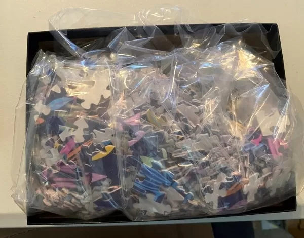
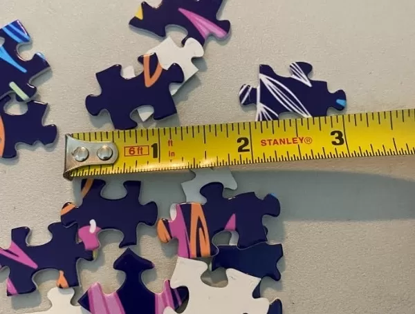
Jigsaw Puzzle (520 Pieces)
Discover the wonders of biology with our Media Storehouse Animal Cell Jigsaw Puzzle! Featuring an intricately detailed diagram of an animal cell from Science Photo Library, this educational puzzle is not only fun but also an excellent way to learn about the complex structures and functions that make up the fundamental unit of life. Ideal for individuals of all ages, this challenging yet rewarding puzzle will help you gain a deeper understanding of cellular anatomy while providing hours of engaging entertainment. Assemble the pieces to reveal the intricacies of the animal cell's internal and external structures, including the nucleus, mitochondria, ribosomes, and more. Perfect for homeschooling, classrooms, or just for personal enrichment, our Animal Cell Jigsaw Puzzle is a must-have for anyone interested in the marvels of biology. So why wait? Dive into the microscopic world and start piecing together the mysteries of the animal cell today!
Made in the USA, 520-piece puzzles measure 16" x 20" (40.6 x 50.8 cm). Every puzzle is meticulously printed on glossy photo paper, which has a strong 1.33 mm thickness. Delivered in a black storage cardboard box, these puzzles are both stylish and practical. (Note: puzzles contain small parts and are not suitable for children under 3 years of age.)
Jigsaw Puzzles are an ideal gift for any occasion
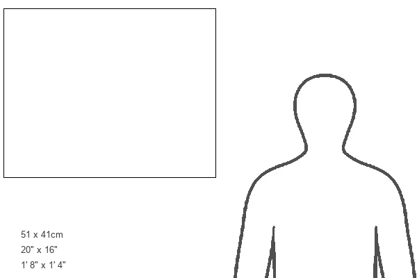
Estimated Product Size is 50.8cm x 40.5cm (20" x 15.9")
These are individually made so all sizes are approximate
Artwork printed orientated as per the preview above, with landscape (horizontal) or portrait (vertical) orientation to match the source image.
EDITORS COMMENTS
This print from Science Photo Library showcases the intricate anatomy of an animal cell. With meticulous detail, the diagram presents both the internal and external features of this essential building block of life. The artwork beautifully captures the complexity and elegance found within a single cut-away view. The image allows us to explore the fascinating world inside an animal cell, revealing its various organelles and structures that play crucial roles in cellular function. From the nucleus, which houses genetic material and controls cellular activities, to mitochondria responsible for energy production, each component is depicted with precision. Highlighted in vibrant colors against a dark background, we can observe key elements such as ribosomes involved in protein synthesis, endoplasmic reticulum facilitating transportation within cells, lysosomes responsible for waste disposal, and plasma membrane acting as a protective barrier. This illustration serves as a visual feast for biology enthusiasts or anyone seeking to understand the intricacies of life at a microscopic level. It exemplifies how art can merge seamlessly with science to create educational masterpieces that inspire curiosity and appreciation for our biological existence.
MADE IN THE USA
Safe Shipping with 30 Day Money Back Guarantee
FREE PERSONALISATION*
We are proud to offer a range of customisation features including Personalised Captions, Color Filters and Picture Zoom Tools
SECURE PAYMENTS
We happily accept a wide range of payment options so you can pay for the things you need in the way that is most convenient for you
* Options may vary by product and licensing agreement. Zoomed Pictures can be adjusted in the Cart.


![(15) [Gate of all Nations, Persepolis, Fars], 1840s-60s. Creator: Luigi Pesce](/sq/731/15-gate-all-nations-persepolis-fars-20171901.jpg.webp)