Beta Sheet Collection (#11)

The beta sheet, a crucial element in the world of molecular biology and protein structure, plays a significant role in various biological processes

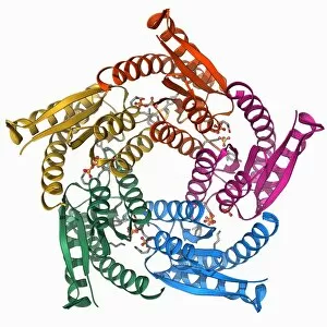
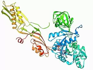
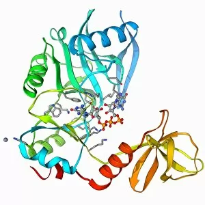
Background imageBeta Sheet Collection: Nucleotide exchange factor F006 / 9292

Nucleotide exchange factor F006 / 9292
Nucleotide exchange factor. Molecular model of the nucleotide exchange factor protein GrpE complexed with the chaperone protein DnaK

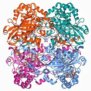
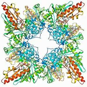
Background imageBeta Sheet Collection: Lumazine synthase molecule F006 / 9291

Lumazine synthase molecule F006 / 9291
Lumazine synthase molecule. Molecular model showing the structure of a lumazine synthase enzyme molecule from a Brucella abortus bacterium

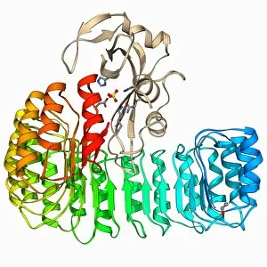
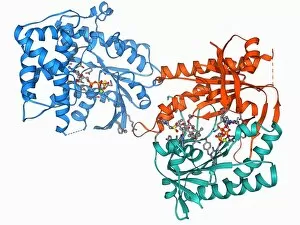
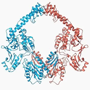
Background imageBeta Sheet Collection: NAD-dependent DNA ligase molecule F006 / 9290

NAD-dependent DNA ligase molecule F006 / 9290
NAD dependent DNA ligase. Molecular model of NAD(+)-dependent DNA ligase. DNA ligase is an enzyme that binds two strands of DNA (deoxyribonucleic acid) together

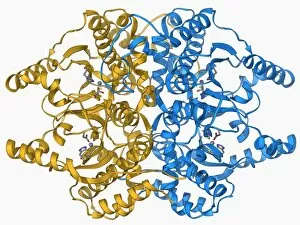
Background imageBeta Sheet Collection: Human catalsae, molecular model F006 / 9288

Human catalsae, molecular model F006 / 9288
Human catalase, molecular model. This enzyme catalyses the break down of hydrogen peroxide to water and oxygen. Hydrogen peroxide is a highly toxic byproduct of a number of normal cellular processes

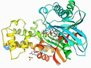
Background imageBeta Sheet Collection: Beta-carbonic anhydrase molecule F006 / 9286

Beta-carbonic anhydrase molecule F006 / 9286
Beta-carbonic anhydrase, molecular model. This enzyme is from the red algae Porphyridium purpureum. It catalyses the reversible hydration of carbon dioxide

Background imageBeta Sheet Collection: Ribonuclease bound to inhibitor F006 / 9287

Ribonuclease bound to inhibitor F006 / 9287
Ribonuclease bound to inhibitor, molecular model. Ribonuclease (RNase) is a type of nuclease that catalyses the degradation of RNA (ribonucleic acid)

Background imageBeta Sheet Collection: Elongation factor G F006 / 9284

Elongation factor G F006 / 9284
Elongation factor G. Molecular model of elongation factor G (EF-G) complexed with GDP (guanosine diphosphate). This enzyme is involved in the elongation of polypeptide chains during translation
Background imageBeta Sheet Collection: DNA Holliday junction, molecular model F006 / 9285

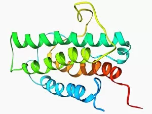
DNA Holliday junction, molecular model F006 / 9285
DNA Holliday junction. Molecular model of a Holliday junction (centre) between homologous strands of DNA (deoxyribonucleic acid)
Background imageBeta Sheet Collection: Coagulation factor complex molecule F006 / 9283

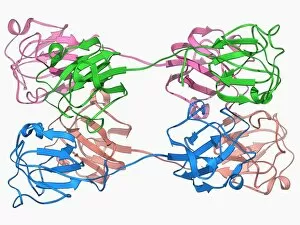
Coagulation factor complex molecule F006 / 9283
Coagulation factor complex molecule. Molecular model showing a blood clotting factor VIIa-tissue factor complex molecule. Tissue factor, also known as factor III

Background imageBeta Sheet Collection: Yeast DNA recognition, molecular model F006 / 9282

Yeast DNA recognition, molecular model F006 / 9282
Yeast DNA recognition. Computer model showing a GAL4 transcription activator protein bound to a yeast DNA (deoxyribonucleic acid) molecule (red and blue)

Background imageBeta Sheet Collection: Creatine kinase molecule F006 / 9276

Creatine kinase molecule F006 / 9276
Creatine kinase molecule. This enzyme catalyses the phosphorylation of creatine to creatine phosphate. It plays an important role in energy metabolism in cells with high or fluctuating energy

Background imageBeta Sheet Collection: Human transferrin receptor molecule F006 / 9280

Human transferrin receptor molecule F006 / 9280
Human transferrin receptor, molecular model. This molecule is found on the surface of a cell. It binds transferrins, iron-binding glycoproteins found in the blood plasma

Background imageBeta Sheet Collection: Sex hormone-binding globulin molecule F006 / 9281

Sex hormone-binding globulin molecule F006 / 9281
Sex hormone-binding globulin. Molecular model of the sex hormone-binding globulin (SHBG) protein complexed with the male sex hormone dihydrotestosterone

Background imageBeta Sheet Collection: Adenylyl cyclase enzyme molecule F006 / 9279

Adenylyl cyclase enzyme molecule F006 / 9279
Adenylyl cyclase. Molecular model of adenylyl cyclase complexed with an inhibitor. This enzyme catalyses the conversion of ATP (adenosine triphosphate) to cyclic AMP (cAMP) and pyrophosphate

Background imageBeta Sheet Collection: Citrate synthase molecule F006 / 9277

Citrate synthase molecule F006 / 9277
Citrate synthase, molecular model. This enzyme is involved in the first step of the citric acid (or Krebs) cycle, the process by which mitochondria convert glucose to energy

Background imageBeta Sheet Collection: Cobra venom molecule F006 / 9278

Cobra venom molecule F006 / 9278
Cobra venom. Molecular model of an alpha-cobratoxin (snake venom protein) from an Indochinese spitting cobra (Naja siamensis)

Background imageBeta Sheet Collection: DNA clamp complexed with DNA molecule F006 / 9274

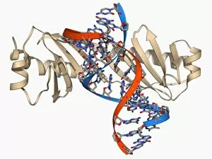
DNA clamp complexed with DNA molecule F006 / 9274
DNA clamp complexed with DNA molecule. Molecular model showing a sliding DNA (deoxyribonucleic acid) clamp (beige) complexed with a molecule of DNA (red and blue)

Background imageBeta Sheet Collection: Synthetic triple helical peptide molecule F006 / 9275

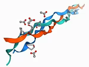
Synthetic triple helical peptide molecule F006 / 9275
Synthetic triple helical peptide, molecular model

Background imageBeta Sheet Collection: mRNA capping enzyme molecule F006 / 9273

mRNA capping enzyme molecule F006 / 9273
mRNA capping enzyme. Molecular model of the mRNA capping enzyme mRNA guanylyltransferase complexed with a cap analogue

Background imageBeta Sheet Collection: Adenylyl cyclase enzyme molecule F006 / 9271

Adenylyl cyclase enzyme molecule F006 / 9271
Adenylyl cyclase enzyme, molecular model. This enzyme catalyses the conversion of ATP (adenosine triphosphate) to cyclic AMP (cAMP) and pyrophosphate. cAMP regulates numerous cell functions

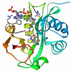
Background imageBeta Sheet Collection: Serotonin N-acetyl transferase molecule F006 / 9272

Serotonin N-acetyl transferase molecule F006 / 9272
Serotonin N-acetyl transferase. Molecular model of serotonin N-acetyl transferase complexed with coenzyme A-S-acetyltryptamine

Background imageBeta Sheet Collection: HGPRTase molecule F006 / 9270

HGPRTase molecule F006 / 9270
HGPRTase. Molecular model of hypoxanthine-guanine phosphoribosyltransferase (HGPRTase) from the malaria parasite Plasmodium falciparum. HGPRTase is involved in the purine salvage pathway

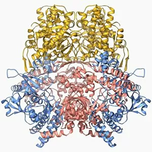
Background imageBeta Sheet Collection: Bleomycin hydrolase molecule F006 / 9265

Bleomycin hydrolase molecule F006 / 9265
Bleomycin hydrolase. Molecular model of the cysteine protease bleomycin hydrolase. This enzyme is thought to be a major cause of resistance to the bleomycin chemotherapy drug

Background imageBeta Sheet Collection: Gene activator protein F006 / 9269

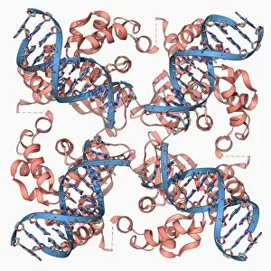
Gene activator protein F006 / 9269
Gene activator protein. Molecular model of catabolite gene activator protein (CAP, pink and green) bound to a molecule of deoxyribonucleic acid (DNA, across top)

Background imageBeta Sheet Collection: TATA box-binding protein and DNA F006 / 9267

TATA box-binding protein and DNA F006 / 9267
TATA box-binding protein and DNA. Molecular model showing a TATA box-binding protein (TBP) complexed with a strand of DNA (deoxyribonucleic acid, red and blue)

Background imageBeta Sheet Collection: CD4 protein molecule F006 / 9266

CD4 protein molecule F006 / 9266
CD4 protein. Molecular model of the CD4 T cell (T lymphocyte) receptor protein. CD4 binds to the surface proteins on other cells and pathogenic micro-organisms

Background imageBeta Sheet Collection: Calcium-binding protein molecule F006 / 9268

Calcium-binding protein molecule F006 / 9268
Calcium-binding protein. Molecule model of the calcium-binding protein calmodulin (CaM). This protein is found in all eukaryotic cells

Background imageBeta Sheet Collection: E coli Holliday junction complex F006 / 9261

E coli Holliday junction complex F006 / 9261
E. coli Holliday junction complex. Molecular model of a RuvA protein (red) in complex with a Holliday junction between homologous strands of DNA (deoxyribonucleic acid, blue) from an E

Background imageBeta Sheet Collection: Metabolic enzyme molecule F006 / 9262

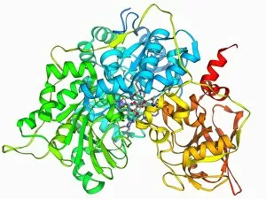
Metabolic enzyme molecule F006 / 9262
Metabolic enzyme. Molecular model of the enzyme aconitase, which is involved in the citric acid (or Krebs) cycle. The citric acid cycle is the process by which mitochondria convert glucose to energy

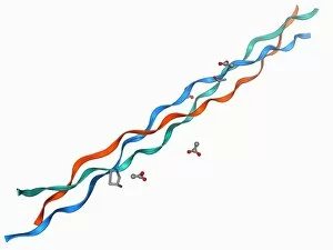
Background imageBeta Sheet Collection: Collagen-like molecule F006 / 9264

Collagen-like molecule F006 / 9264
Collagen-like molecule. Molecular model showing the triple helical structure of a collagen-like molecule

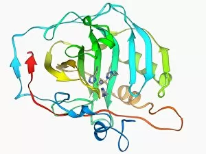
Background imageBeta Sheet Collection: Carbonic anhydrase II molecule F006 / 9263

Carbonic anhydrase II molecule F006 / 9263
Carbonic anhydrase II, molecular model. This enzyme catalyses the reversible hydration of carbon dioxide

Background imageBeta Sheet Collection: Bacteriorhodopsin protein F006 / 9260

Bacteriorhodopsin protein F006 / 9260
Bacteriorhodopsin protein. Molecular model showing the structure of bacteriorhodopsin (bR), a protein found in primitive micro-organisms known as Archaea. This protein acts as a proton pump

Background imageBeta Sheet Collection: ATP synthase molecule F006 / 9258

ATP synthase molecule F006 / 9258
ATP synthase molecule. Molecular model showing the structure of ATP synthase (ATPase) subunit A and C. ATPase is an important enzyme that provides energy for cells through the synthesis of adenosine

Background imageBeta Sheet Collection: Diels-Alder antibody catalyst molecule F006 / 9259

Diels-Alder antibody catalyst molecule F006 / 9259
Diels-Alder antibody catalyst. Molecular model of an antibody that can be used to catalyse the Diels-Alder reaction

Background imageBeta Sheet Collection: Bioluminescent enzyme molecule F006 / 9256

Bioluminescent enzyme molecule F006 / 9256
Bioluminescent enzyme. Molecular model of the bioluminescent enzyme luciferase from the marine bacteria Vibrio harveyi

Background imageBeta Sheet Collection: Myosin molecule F006 / 9255

Myosin molecule F006 / 9255
Myosin. Molecular molecule of a smooth muscle myosin. Myosins are a large family of motor proteins that are responsible for muscle contraction in eukaryotic tissues

Background imageBeta Sheet Collection: Erythropoietin hormone molecule F006 / 9257

Erythropoietin hormone molecule F006 / 9257
Erythropoietin. Molecular model of the human erythropoietin (EPO) hormone. Erythropoietin regulates blood oxygen levels in the body

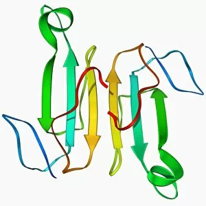
Background imageBeta Sheet Collection: Eye lens protein molecule F006 / 9253

Eye lens protein molecule F006 / 9253
Eye lens protein. Molecular model of beta-crystallin, a protein found in the lens of the eye. The regular arrangement of the protein in the lens is thought to be responsible for its transparency

Background imageBeta Sheet Collection: Beta-galactosidase molecule F006 / 9250

Beta-galactosidase molecule F006 / 9250
Beta-galactosidase, molecular model. This enzyme breaks down sugars containing galactose, such as lactose, into their basic units (monosaccharides)

Background imageBeta Sheet Collection: Type II topoisomerase molecule F006 / 9251

Type II topoisomerase molecule F006 / 9251
Type II topoisomerase, molecular model. The topoisomerase enzymes assist in uncoiling DNA (deoxyribonucleic acid). DNA is usually stored in a supercoiled form

Background imageBeta Sheet Collection: Human tissue factor molecule F006 / 9254

Human tissue factor molecule F006 / 9254
Human tissue factor. Molecular model of human tissue factor, also known as factor III. Tissue factor is found on the outside of blood vessels and plays a role in the coagulation cascade

Background imageBeta Sheet Collection: Nerve growth factor molecule F006 / 9249

Nerve growth factor molecule F006 / 9249
Nerve growth factor (NGF), molecular model. NGF is a small protein, which is involved in the growth, differentiation (specialisation of cells) and survival of particular target neurones (nerve cells)

Background imageBeta Sheet Collection: Collagen-like molecule T3-785 F006 / 9252

Collagen-like molecule T3-785 F006 / 9252
Collagen-like molecule. Molecular model showing the triple helical structure of the collagen-like molecule T3-785

Background imageBeta Sheet Collection: Hexokinase and glucose complex F006 / 9248

Hexokinase and glucose complex F006 / 9248
Hexokinase and glucose complex. Molecular model of a complex between the enzyme hexokinase and the sugar glucose. Hexokinase promotes the conversion (phosphorylation)

Background imageBeta Sheet Collection: T cell receptor B7 molecule F006 / 9247

T cell receptor B7 molecule F006 / 9247
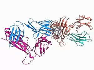
T cell receptor B7. Molecular model of the T cell receptor B7 bound to the viral Tax peptide and MHC class I molecule HLA-A2

Background imageBeta Sheet Collection: Myosin fragment molecule F006 / 9245

Myosin fragment molecule F006 / 9245
Myosin fragment. Molecular molecule of a fragment of striated muscle myosin complexed with ADP (adenosine diphosphate). Myosins are a large family of motor proteins that are responsible for muscle

Background imageBeta Sheet Collection: Scorpion toxin molecule F006 / 9246

Scorpion toxin molecule F006 / 9246
Scorpion toxin. Molecular model of the neurotoxin BJXTR-IT from the Hottentotta judaicus scorpion. This toxin functions by modulating sodium channels

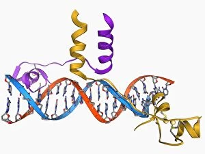
Background imageBeta Sheet Collection: Pit-1 transcription factor bound to DNA F006 / 9242

Pit-1 transcription factor bound to DNA F006 / 9242
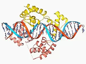
Pit-1 transcription factor bound to DNA. Molecular model showing pituitary-specific positive transcription factor 1 (Pit-1) (yellow and pink) bound to a strand of DNA (deoxyribonucleic acid)


For sale as Licensed Images

Choose your image, Select your licence and Download the media

The beta sheet, a crucial element in the world of molecular biology and protein structure, plays a significant role in various biological processes. From DNA transcription to HIV reverse transcription enzyme, this secondary structure of proteins is an intricate masterpiece. In the realm of DNA transcription, the beta sheet takes center stage as it assists in unraveling genetic information. Its molecular model showcases its complexity and elegance, resembling an artistic creation that captivates scientists worldwide. Not only does the beta sheet contribute to DNA transcription, but it also plays a vital role in combating diseases. The Hepatitis C virus enzyme's molecular model reveals how this secondary structure aids in understanding and potentially treating this infectious disease. Another remarkable example lies within Manganese superoxide dismutase enzyme F006 / 9423. This intricate arrangement highlights how the beta sheet contributes to antioxidant defense mechanisms within our bodies. Moreover, Argonaute protein molecule F006 / 9526 demonstrates how the beta sheet collaborates with microRNA to regulate gene expression. This interaction holds immense potential for therapeutic interventions targeting various diseases. Immunoglobulin G antibody and egg white F006 / 9682 showcase yet another fascinating aspect of the beta sheet's versatility. Here we witness its involvement in immune responses against foreign substances present within our bodies. Furthermore, Cytochrome P450 complex F006 / 9669 illustrates how this secondary structure enables drug metabolism and detoxification processes essential for maintaining human health. Succinyl-CoA synthetase enzyme F006 / 9592 emphasizes another critical function of the beta sheet: energy production through cellular respiration pathways. Its presence ensures efficient conversion of succinyl-CoA into ATP molecules necessary for sustaining life processes. Additionally, RNA-induced silencing complex F006 / 9586 unveils how the beta sheet participates in gene regulation by suppressing specific mRNA molecules through small interfering RNAs (siRNAs).