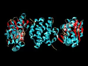
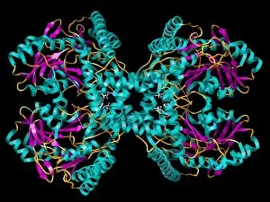
Beta Sheet Collection (#13)

The beta sheet, a crucial element in the world of molecular biology and protein structure, plays a significant role in various biological processes

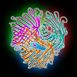
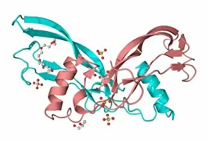
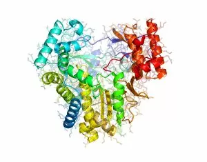
Background imageBeta Sheet Collection: Sucrose-specific porin molecule C013 / 8870

Sucrose-specific porin molecule C013 / 8870
Sucrose-specific porin molecule. Moleuclar model showing the secondary and quaternary structure of a molecule of sucrose-specific porin

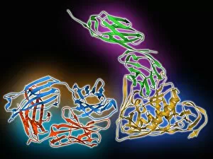
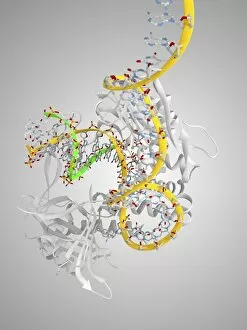
Background imageBeta Sheet Collection: HIV antibody therapy, molecular model C013 / 7908

HIV antibody therapy, molecular model C013 / 7908
HIV antibody therapy. Molecular model of the interaction of the HIV surface protein gp120 (yellow, bottom right) as it interacts with a human white blood cell surface protein (CD4, green)

Background imageBeta Sheet Collection: Glycated haemoglobin molecule C013 / 7781

Glycated haemoglobin molecule C013 / 7781
Glycated haemoglobin molecule. Computer model of a glycated haemoglobin molecule. The alpha and beta subunits of the haemoglobin are blue and pink, and the iron-containing haem groups are grey

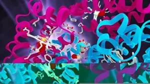
Background imageBeta Sheet Collection: Glycated haemoglobin molecule C013 / 7779

Glycated haemoglobin molecule C013 / 7779
Glycated haemoglobin molecule. Computer model showing a glucose molecule (centre) bound to a molecule of haemoglobin. The alpha and beta subunits of the haemoglobin are blue and pink

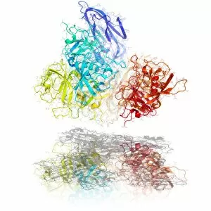
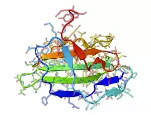
Background imageBeta Sheet Collection: Norwalk virus capsid protein

Norwalk virus capsid protein. Molecular model showing the secondary structure of the capsid protein from a norwalk virus particle

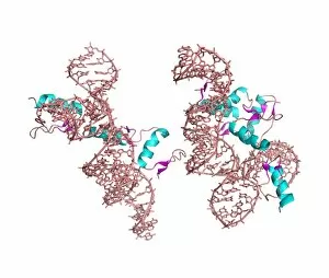
Background imageBeta Sheet Collection: Zinc finger-RNA complex

Zinc finger-RNA complex. Computer models of zinc finger protein molecules complexed with RNA (ribonucleic acid) molecules

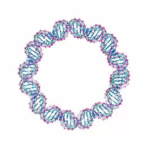
Background imageBeta Sheet Collection: DNA loop, molecular model

DNA loop, molecular model
DNA loop. Theoretical molecular model of a loop structure for DNA in its chromatin form. DNA (deoxyribonucleic acid) is a helical molecule (here the helix itself forms a circular loop)

Background imageBeta Sheet Collection: RNA polymerase II molecule

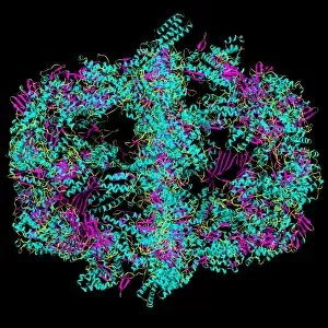
RNA polymerase II molecule
RNA polymerase II. Computer model showing the secondary structure of the enzyme RNA polymerase II. The molecule comprises 12 subunits

Background imageBeta Sheet Collection: Protein structure, artwork

Protein structure, artwork
Protein structure. Computer artwork of alpha helices (coils) and beta sheets (ribbons) of the secondary structure of a protein

Background imageBeta Sheet Collection: PPAR regulatory molecule

PPAR regulatory molecule. Molecular model of peroxisome proliferator-activated receptor gamma (PPARG, pink) bound to a DNA (deoxyribonucleic acid) molecule (orange and green)

Background imageBeta Sheet Collection: Viral DNA polymerase in complex with DNA

Viral DNA polymerase in complex with DNA. Computer model showing the active site of a phi29 DNA polymerase molecule (grey ribbons) in complex with DNA (deoxyribonucleic acid, yellow)

Background imageBeta Sheet Collection: Tubulin dimer, molecular model

Tubulin dimer, molecular model
Tubulin. Molecular model of a tubulin dimer. The beta-tubulin subunit (gold) is at top and the alpha-tubulin subunit (green) is at bottom

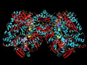
Background imageBeta Sheet Collection: Xanthine dehydrogenase molecule

Xanthine dehydrogenase molecule. Computer model showing the molecular structure of the Xanthine dehydrogenase (XDH) enzyme

Background imageBeta Sheet Collection: Iron-hydrogenase molecule

Iron-hydrogenase molecule. Computer model showing the molecular structure of an iron-hydrogenase ([FE]-hydrogenase) enzyme from the Methanocaldococcus jannaschii archaeon

Background imageBeta Sheet Collection: Fatty acid synthase in complex with NADP+

Fatty acid synthase in complex with NADP+. Computer model showing the molecular structure of fatty acid synthase (FAS) complexed with nicotinamide adenine dinucleotide phosphate (NADP+)

Background imageBeta Sheet Collection: Feline distemper virus particle

Feline distemper virus particle. Molecular model showing the structure of the capsid (outer protein coat) of a feline panleukopenia virus particle. This parovirus causes distemper in cats

Background imageBeta Sheet Collection: Cholesterol producing enzyme and statin

Cholesterol producing enzyme and statin. Computer model showing the molecular structure of HMG-CoA reductase (HMGCR) in complex with Atorvastatin

Background imageBeta Sheet Collection: Tobacco mosaic virus, molecular model

Tobacco mosaic virus, molecular model
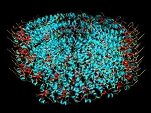
Tobacco mosaic virus. Computer model showing the molecular structure of a tobacco mosaic virus (TMV) particle (virion). This plant virus is a tobamovirus, and has an RNA (ribonucleic acid) genome

Background imageBeta Sheet Collection: Selenocysteine synthase enzyme molecule

Selenocysteine synthase enzyme molecule. Computer model showing the molecular structure of the enzyme selenocysteine synthase (SecS)
Background imageBeta Sheet Collection: Spanish flu H1 antigen, molecular model

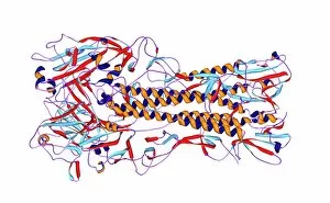
Spanish flu H1 antigen, molecular model
Spanish flu virus antigen. Computer model showing the molecular structure of a haemagglutinin 1 (H1) antigen from the Spanish influenza (flu) virus particle

Background imageBeta Sheet Collection: Major sperm protein molecule

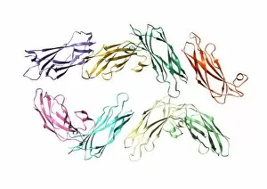
Major sperm protein molecule
Major sperm protein. Computer model showing the secondary structure of the peptide chains that make up major sperm protein (MSP)

Background imageBeta Sheet Collection: Proteinase inhibitor molecule

Proteinase inhibitor molecule
Proteinase inhibitor. Molecular model showing the secondary structure of alpha-1 proteinase inhibitor, also known as alpha 1-antitrypsin

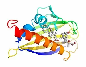
Background imageBeta Sheet Collection: Insulin receptor substrate 1 molecule

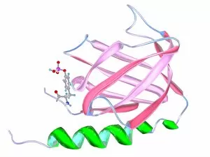
Insulin receptor substrate 1 molecule
Insulin receptor substrate 1. Molecular model of the secondary structure of the protein insulin receptor substrate 1 (IRS1), bound to a molecule of phosphotyrosine (ball and stick molecule)

Background imageBeta Sheet Collection: Inhibin beta A molecule

Inhibin beta A molecule. Computer model showing the crystal structure of a molecule of the protein Inhibin beta A (INHBA)

Background imageBeta Sheet Collection: Thrombopoietin hormone molecule

Thrombopoietin hormone molecule. Computer model showing the secondary structure of a molecule of the hormone thrombopoietin (TPO)

Background imageBeta Sheet Collection: Renin and inhibitor complex

Renin and inhibitor complex. Computer model showing the secondary structure of the enzyme renin complexed with inhibitor 7

Background imageBeta Sheet Collection: Human chorionic gonadotrophin molecule

Human chorionic gonadotrophin molecule. Computer model showing the crystal structure of a molecule of the hormone human chorionic gonadotrophin (hCG)

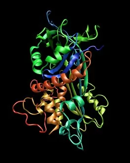
Background imageBeta Sheet Collection: Kinase molecule, secondary structure

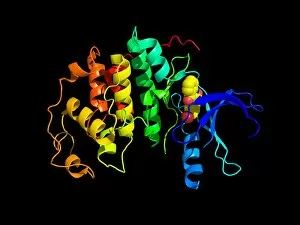
Kinase molecule, secondary structure
Kinase molecule, computer model. Kinases are enzymes that catalyse the transfer of phosphate groups from a high-energy phosphate-containing molecule (such as ATP or ADP)

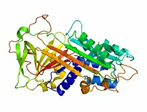
Background imageBeta Sheet Collection: Lipase molecule, secondary structure

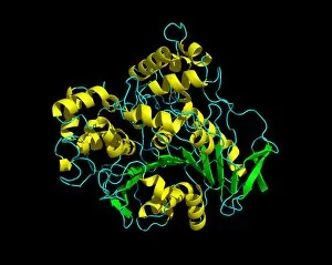
Lipase molecule, secondary structure
Lipase molecule, computer model. Lipase is an enzyme that breaks lipids (fats) into fatty acids and glycerol. Human pancreatic lipase is the main enzyme responsible for breaking down fat in the human

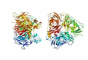
Background imageBeta Sheet Collection: Ferroxidase enzyme, molecular model

Ferroxidase enzyme, molecular model
Ferroxidase enzyme. Molecular model showing two views of the secondary structure of the human enzyme ferroxidase, also known as ceruloplasmin. Copper atoms are represented as red spheres

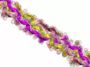
Background imageBeta Sheet Collection: Synthetic peptide fibre, molecular model

Synthetic peptide fibre, molecular model
Synthetic peptide fibre. Molecular model of a synthetic collagen-like peptide fibre, showing three different ways of representing the structure. Peptides are small molecules formed from amino acids

Background imageBeta Sheet Collection: Viral dUTPase enzyme, molecular model

Viral dUTPase enzyme, molecular model
dUTPase enzyme. Molecular model of the enzyme dUTP pyrophosphatase (dUTPase) from the feline immunodeficiency virus (FIV)

Background imageBeta Sheet Collection: Cyclin-depenent kinase 5 molecule

Cyclin-depenent kinase 5 molecule
Cyclin-dependent kinase 5. Molecular model showing the secondary structure of the enzyme cylcin- dependent kinase 5 (cdk5)

Background imageBeta Sheet Collection: Bacteriochlorophyll-containing protein

Bacteriochlorophyll-containing protein. Molecular models showing two views of the bacteriochlorophyll-containing protein found in the bacterium Prosthecochloris aestuarii

Background imageBeta Sheet Collection: Antithrombin molecule

Antithrombin molecule
Antithrombin, molecular model. Antithrombin is a glycoprotein produced in the liver that inhibits enzymes involved in blood clotting

Background imageBeta Sheet Collection: Interleukin-12 protein molecule

Interleukin-12 protein molecule. Molecular model of a molecule of the cytokine protein interleukin-12 (IL-12). The secondary structure of the molecule is seen here

Background imageBeta Sheet Collection: Microglobulin protein, molecular model

Microglobulin protein, molecular model
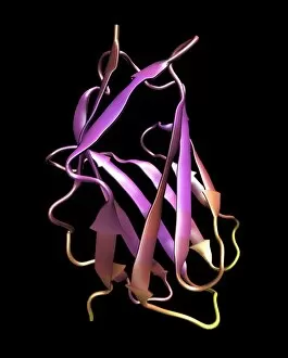
Microglobulin protein. Molecular models of two overlapping views of the protein beta-2 microglobulin. This protein, with a relatively small molecular mass

Background imageBeta Sheet Collection: Birch pollen allergen

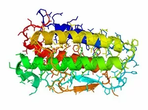
Birch pollen allergen. Molecular model of the secondary structure of Bet v 1l, the molecule responsible for allergic reactions to birch pollen

Background imageBeta Sheet Collection: Cytochrome P450 molecule

Cytochrome P450 molecule
Cytochrome P450 protein. Molecular model showing the secondary structure of cytochrome P450. This protein plays a crucial role in metabolism in animals (including humans), fungi, plants and bacteria

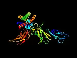
Background imageBeta Sheet Collection: Cd28 antigen molecule

Cd28 antigen molecule. Computer model showing the secondary structure of cd28. Cd28 is an antigen found on the surface of T cells

Background imageBeta Sheet Collection: Mouse urinary protein, molecular model

Mouse urinary protein, molecular model
Mouse urinary protein (MUP), molecular model. MUP refers to a family of similar proteins found in mouse urine. The proteins act as pheromones, airborne chemical signals

Background imageBeta Sheet Collection: Bluetongue virus protein structure

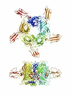
Bluetongue virus protein structure. Molecular model of some of the protein structures found in the protein coating of the bluetongue virus (BTV)

Background imageBeta Sheet Collection: Erythropoietin hormone

Erythropoietin hormone
Erythropoietin. Computer model of a molecule of the hormone erythropoietin. This is a glycoprotein that consists of a single-chain polypeptide of 165 amino acids

Background imageBeta Sheet Collection: Testosterone bound to receptor, artwork

Testosterone bound to receptor, artwork
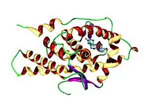
Testosterone bound to receptor. Molecular model of a molecule of the male sex hormone testosterone (ball-and-stick primary structure) bound to a human androgen receptor (secondary structure)

Background imageBeta Sheet Collection: Green fluorescent protein, computer model

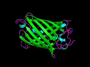
Green fluorescent protein, computer model. This protein is found in the jellyfish Aequorea victoria. When ultraviolet or blue light shines on the protein, it emits green light

Background imageBeta Sheet Collection: Cobra venom action, molecular model

Cobra venom action, molecular model
Cobra (Naja sp.) venom action. Molecular model showing top (upper centre) and side (lower centre) views of the secondary structure of an alpha-cobratoxin (snake venom protein)

Background imageBeta Sheet Collection: RNA polymerase from Norwalk virus

RNA polymerase from Norwalk virus
RNA polymerase enzyme from Norwalk virus, molecular model. This enzyme makes RNA (ribonucleic acid) from the RNA template within the virus

Background imageBeta Sheet Collection: TGF beta, molecular model

TGF beta, molecular model
TGF beta molecule. Molecular model showing the primary (rods) and secondary structure (arrows) of transforming growth factor beta (TGF beta)


For sale as Licensed Images

Choose your image, Select your licence and Download the media

The beta sheet, a crucial element in the world of molecular biology and protein structure, plays a significant role in various biological processes. From DNA transcription to HIV reverse transcription enzyme, this secondary structure of proteins is an intricate masterpiece. In the realm of DNA transcription, the beta sheet takes center stage as it assists in unraveling genetic information. Its molecular model showcases its complexity and elegance, resembling an artistic creation that captivates scientists worldwide. Not only does the beta sheet contribute to DNA transcription, but it also plays a vital role in combating diseases. The Hepatitis C virus enzyme's molecular model reveals how this secondary structure aids in understanding and potentially treating this infectious disease. Another remarkable example lies within Manganese superoxide dismutase enzyme F006 / 9423. This intricate arrangement highlights how the beta sheet contributes to antioxidant defense mechanisms within our bodies. Moreover, Argonaute protein molecule F006 / 9526 demonstrates how the beta sheet collaborates with microRNA to regulate gene expression. This interaction holds immense potential for therapeutic interventions targeting various diseases. Immunoglobulin G antibody and egg white F006 / 9682 showcase yet another fascinating aspect of the beta sheet's versatility. Here we witness its involvement in immune responses against foreign substances present within our bodies. Furthermore, Cytochrome P450 complex F006 / 9669 illustrates how this secondary structure enables drug metabolism and detoxification processes essential for maintaining human health. Succinyl-CoA synthetase enzyme F006 / 9592 emphasizes another critical function of the beta sheet: energy production through cellular respiration pathways. Its presence ensures efficient conversion of succinyl-CoA into ATP molecules necessary for sustaining life processes. Additionally, RNA-induced silencing complex F006 / 9586 unveils how the beta sheet participates in gene regulation by suppressing specific mRNA molecules through small interfering RNAs (siRNAs).