Beta Sheet Collection (#10)

The beta sheet, a crucial element in the world of molecular biology and protein structure, plays a significant role in various biological processes

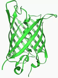
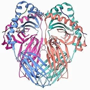
Background imageBeta Sheet Collection: Green fluorescent protein molecule F006 / 9343

Green fluorescent protein molecule F006 / 9343
Green fluorescent protein (GFP), molecular model. The molecule has a cylindrical structure formed from beta sheets (ribbons). GFP is found in the Pacific jellyfish Aequorea victoria
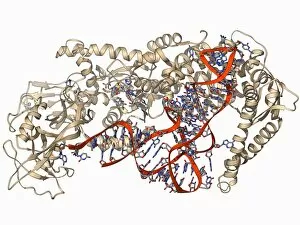
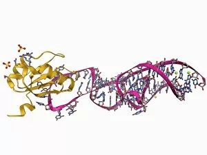
Background imageBeta Sheet Collection: Valyl-tRNA synthetase molecule F006 / 9342

Valyl-tRNA synthetase molecule F006 / 9342
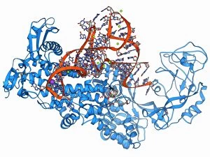
Valyl-tRNA synthetase protein molecule. Molecular model showing bacterial valyl-tRNA synthetase complexed with valyl tRNA (transfer ribonucleic acid)

Background imageBeta Sheet Collection: Xanthine dehydrogenase molecule F006 / 9336

Xanthine dehydrogenase molecule F006 / 9336
Xanthine dehydrogenase molecule. Molecular model of the Xanthine dehydrogenase (XDH) enzyme. XDH is an oxidoreductase enzyme that catalyses the last two steps in the formation of urate (uric acid)

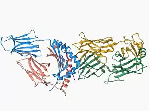
Background imageBeta Sheet Collection: T cell receptor antigen complex F006 / 9339

T cell receptor antigen complex F006 / 9339
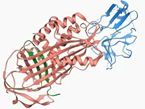
T cell receptor antigen complex. Molecular model of the alphabeta T cell receptor bound to the influenza haemagglutinin antigen and MHC class II molecule HLA-DR1

Background imageBeta Sheet Collection: Transcription factor complexed with DNA F006 / 9337

Transcription factor complexed with DNA F006 / 9337
Transcription factor complexed with DNA. Molecular model showing the transcriptional factor c-fos c-jun (yellow and pink) bound to a strand of DNA (deoxyribonucleic acid, orange and blue)

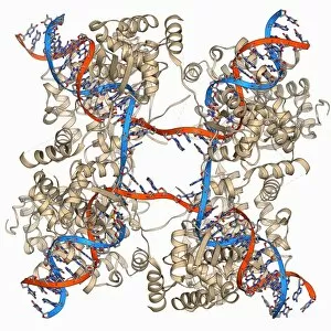
Background imageBeta Sheet Collection: DNA Holliday junction complex F006 / 9334

DNA Holliday junction complex F006 / 9334
DNA Holliday junction complex. Molecular model of the enzyme FLP recombinase in complex with a Holliday junction between homologous strands of DNA (deoxyribonucleic acid)

Background imageBeta Sheet Collection: Birch pollen allergen F006 / 9335

Birch pollen allergen F006 / 9335
Birch pollen allergen. Molecular model of Bet v 1l, the molecule responsible for allergic reactions to birch pollen. Here

Background imageBeta Sheet Collection: Iron storage molecule F006 / 9331

Iron storage molecule F006 / 9331
Iron storage molecule. Molecular model of ferritin, a protein that acts as an iron store and is mainly found in the liver, kidneys and spleen

Background imageBeta Sheet Collection: Bacterial ribosome, molecular model F006 / 9332

Bacterial ribosome, molecular model F006 / 9332
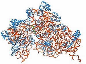
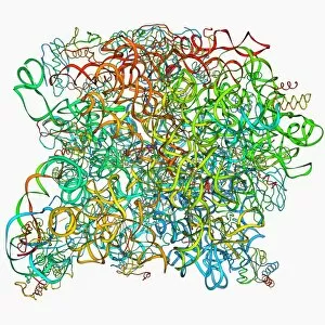
Bacterial ribosome. Molecular model of a 30S (small) ribosomal sub-unit from the bacteria Thermus thermophilus. Ribosomes are composed of protein and RNA (ribonucleic acid)

Background imageBeta Sheet Collection: Beta secretase enzyme, molecular model F006 / 9333

Beta secretase enzyme, molecular model F006 / 9333
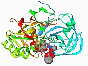
Beta secretase enzyme. Molecular model of the enzyme beta secretase bound to an inhibitor molecule. Beta secretase is a membrane-associated aspartic protease

Background imageBeta Sheet Collection: Archaeon ribosome, molecular model F006 / 9328

Archaeon ribosome, molecular model F006 / 9328
Archaeon ribosome. Molecular model showing the structure of a 50S (large) ribosome from the archaeon Haloarcula marismortui. Ribosomes are composed of protein and RNA (ribonucleic acid)

Background imageBeta Sheet Collection: Isoleucyl-tRNA synthetase molecule F006 / 9329

Isoleucyl-tRNA synthetase molecule F006 / 9329
Isoleucyl-tRNA synthetase protein molecule. Molecular model showing bacterial isoleucyl-tRNA synthetase complexed with aspartyl tRNA (transfer ribonucleic acid)

Background imageBeta Sheet Collection: Interferon gamma molecule and receptor F006 / 9330

Interferon gamma molecule and receptor F006 / 9330
Interferon gamma. Molecular model of the interferon gamma dimer attached to the alpha chain of the interferon gamma receptor

Background imageBeta Sheet Collection: Bacteriorhodopsin protein F006 / 9327

Bacteriorhodopsin protein F006 / 9327
Bacteriorhodopsin protein. Molecular model showing the structure of bacteriorhodopsin (bR), a protein found in primitive micro-organisms known as Archaea. This protein acts as a proton pump

Background imageBeta Sheet Collection: Tetraubiquitin protein molecule F006 / 9326

Tetraubiquitin protein molecule F006 / 9326
Tetraubiquitin protein, molecular model. Ubiquitin is found in all eukaryotic cells. When a protein is damaged or old it will be tagged by several ubiquitin molecules

Background imageBeta Sheet Collection: Nucleosome molecule F006 / 9323

Nucleosome molecule F006 / 9323
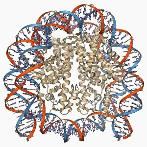
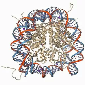
Nucleosome, molecular model. A nucleosome is a subunit of chromatin, the substance that forms chromosomes. It consists of a short length of DNA (deoxyribonucleic acid)

Background imageBeta Sheet Collection: Rhodopsin molecule F006 / 9325

Rhodopsin molecule F006 / 9325
Rhodopsin molecule. Molecular model of the rhodopsin complex. Rhodopsin (also called visual purple) is a pigment found in the rod photoreceptor cells in the retina of the eye

Background imageBeta Sheet Collection: Cre-Lox recombination, molecular model F006 / 9322

Cre-Lox recombination, molecular model F006 / 9322
Cre-Lox recombination. Molecular model of the enzyme CRE (cyclization recombination) recombinase (beige) mediating the recombination of strands of DNA (deoxyribonucleic acid, red and blue)

Background imageBeta Sheet Collection: Proteinase inhibitor molecule F006 / 9321

Proteinase inhibitor molecule F006 / 9321
Proteinase inhibitor. Molecular model of a proteinase inhibitor, or antitrypsin, molecule bound to a tryspin protease. The proteinase inhibitor is a type of serine protease inhibitor (serpin)

Background imageBeta Sheet Collection: Voltage-gated potassium channel F006 / 9324

Voltage-gated potassium channel F006 / 9324
Voltage-gated potassium channel. Molecular model of a voltage-gated potassium (Kv) ion channel. Ion channels are membrane-spanning proteins that form pores in cell membranes

Background imageBeta Sheet Collection: Insect antifreeze protein F006 / 9320

Insect antifreeze protein F006 / 9320
Insect antifreeze protein. Molecular model of an insect antifreeze protein (AFP) from the Tenebrio molitor beetle

Background imageBeta Sheet Collection: Nucleosome molecule F006 / 9314

Nucleosome molecule F006 / 9314
Nucleosome, molecular model. A nucleosome is a subunit of chromatin, the substance that forms chromosomes. It consists of a short length of DNA (deoxyribonucleic acid)

Background imageBeta Sheet Collection: Ebola matrix protein molecule F006 / 9317

Ebola matrix protein molecule F006 / 9317
Ebola matrix protein. Molecular model of the Ebola virus matrix protein VP40. This membrane-associated protein is thought to be necessary for the assembly and budding of viral particles

Background imageBeta Sheet Collection: Insect antifreeze protein F006 / 9319

Insect antifreeze protein F006 / 9319
Insect antifreeze protein. Molecular model of an insect antifreeze protein (AFP) from the spruce budworm (Choristoneura fumiferana)

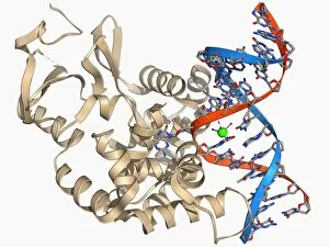
Background imageBeta Sheet Collection: Oxoguanine glycosylase complex F006 / 9318

Oxoguanine glycosylase complex F006 / 9318
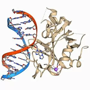
Oxoguanine glycosylase complex. Computer model showing an 8-Oxoguanine glycosylase (OGG1) molecule (beige) bound to a section of DNA (deoxyribonucleic acid, red and blue)

Background imageBeta Sheet Collection: Transcription repressor molecule F006 / 9316

Transcription repressor molecule F006 / 9316
Transcription repressor. Molecular model of the Tup 1 transcription repressor protein. Transcription repressors bind to specific sequences of DNA (deoxyribonucleic acid)

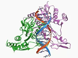
Background imageBeta Sheet Collection: Restriction enzyme and DNA F006 / 9315

Restriction enzyme and DNA F006 / 9315
Restriction enzyme and DNA. Molecular model showing an EcoRI endonuclease enzyme (purple and green) bound to a DNA (deoxyribonucleic acid) molecule (red and blue)

Background imageBeta Sheet Collection: Green fluorescent protein molecule F006 / 9313

Green fluorescent protein molecule F006 / 9313
Green fluorescent protein (GFP), molecular model. The molecule has a cylindrical structure formed from beta sheets (ribbons). GFP is found in the Pacific jellyfish Aequorea victoria

Background imageBeta Sheet Collection: MoaC protein molecule F006 / 9312

MoaC protein molecule F006 / 9312
MoaC protein. Molecular model of the molybdenum cofactor biosynthesis protein MoaC. This enzyme is involved in carbon, nitrogen and sulphur metabolism

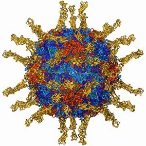
Background imageBeta Sheet Collection: Poliovirus particle F006 / 9306

Poliovirus particle F006 / 9306
Poliovirus particle, molecular model

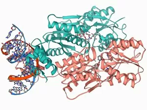
Background imageBeta Sheet Collection: LAC repressor bound to DNA F006 / 9309

LAC repressor bound to DNA F006 / 9309
LAC repressor bound to DNA. Molecular model of a LAC (lactose) repressor molecule (pink and turquoise) interacting with bacterial DNA (deoxyribonucleic acid, red and blue)

Background imageBeta Sheet Collection: Epidermal growth factor molecule F006 / 9311

Epidermal growth factor molecule F006 / 9311
Epidermal growth factor (EGF), molecular model. EGF plays an important role in the regulation of cell growth, proliferation and differentiation

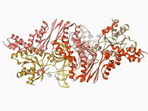
Background imageBeta Sheet Collection: Elongation factors Tu and Ts F006 / 9310

Elongation factors Tu and Ts F006 / 9310
Elongation factors Tu and Ts, molecular model. These enzymes are involved in the elongation of polypeptide chains during translation

Background imageBeta Sheet Collection: Erythropoietin bound to receptors F006 / 9308

Erythropoietin bound to receptors F006 / 9308
Artwork of the human erythropoietin (EPO) hormone molecule (yellow) bound to receptors (pink). Erythropoietin regulates blood oxygen levels in the body

Background imageBeta Sheet Collection: Pyruvate dehydrogenase complex enzyme F006 / 9303

Pyruvate dehydrogenase complex enzyme F006 / 9303
Pyruvate dehydrogenase complex enzyme. Molecular model of dihydrolipoyl transacetylase one of the enzymes in the pyruvate dehydrogenase complex

Background imageBeta Sheet Collection: Oxoguanine glycosylase complex F006 / 9307

Oxoguanine glycosylase complex F006 / 9307
Oxoguanine glycosylase complex. Computer model showing an 8-Oxoguanine glycosylase (OGG1) molecule (beige) bound to a section of DNA (deoxyribonucleic acid, red and blue)

Background imageBeta Sheet Collection: Semliki forest virus capsid F006 / 9297

Semliki forest virus capsid F006 / 9297
Semliki forest virus capsid, molecular model. This virus, named for the forest in Uganda where it was identified, is spread by the bite of mosquitoes. It can infect both humans and animals

Background imageBeta Sheet Collection: ATPase molecule F006 / 9300

ATPase molecule F006 / 9300
ATPase molecule. Molecular model of the central stalk of an ATP synthase (ATPase) molecule from a cow. ATPase is an important enzyme that provides energy for cells through the synthesis of adenosine

Background imageBeta Sheet Collection: Cytochrome P450 and fluconazole F006 / 9302

Cytochrome P450 and fluconazole F006 / 9302
Cytochrome P450 and fluconazole. Molecular model of cytochrome P450 complexed with the anti-fungal compound fluconazole. This protein plays a crucial role in metabolism in animals (including humans)

Background imageBeta Sheet Collection: Bacteriorhodopsin protein F006 / 9299

Bacteriorhodopsin protein F006 / 9299
Bacteriorhodopsin protein. Molecular model showing the structure of bacteriorhodopsin (bR), a protein found in primitive micro-organisms known as Archaea. This protein acts as a proton pump

Background imageBeta Sheet Collection: Human serum albumin molecule F006 / 9301

Human serum albumin molecule F006 / 9301
Human serum albumin, molecular model. Albumin is the most abundant protein in human blood plasma. It is seen here complexed with stearic acid, a saturated fatty acid

Background imageBeta Sheet Collection: HP1 molecule C-terminal domain F006 / 9298

HP1 molecule C-terminal domain F006 / 9298
HP1 molecule C-terminal domain. Molecular model showing the structure of the C terminal (shadow chromo) domain of a heterochromatin protein 1 (HP1) molecule from a mouse

Background imageBeta Sheet Collection: Human poliovirus, molecular model F006 / 9289

Human poliovirus, molecular model F006 / 9289
Human poliovirus particle. Molecular model of the capsid of the human poliovirus. The capsid is a protein coat that encloses the viruss genetic information (genome), stored as RNA (ribonucleic acid)

Background imageBeta Sheet Collection: Green mamba venom molecule F006 / 9296

Green mamba venom molecule F006 / 9296
Green mamba venom. Molecular model of the alpha-dendrotoxin from green mamba (Dendroaspis angusticeps) venom

Background imageBeta Sheet Collection: MHC protein complexed with flu virus F006 / 9294

MHC protein complexed with flu virus F006 / 9294
MHC protein complexed with flu virus. Molecular model showing human class II MHC (major histocompatibility complex) protein HLA-DR1 complexed with an influenza (flu) virus peptide

Background imageBeta Sheet Collection: Hepatitis D virus ribozyme complex F006 / 9295

Hepatitis D virus ribozyme complex F006 / 9295
Hepatitis D virus ribozyme complex. Molecular model showing an RNA (ribonucleic acid) strand from an Hepatitis delta (Hepatitis D) virus genomic ribozyme, complexed with a ribonucleoprotein

Background imageBeta Sheet Collection: Heat shock protein 70 F006 / 9293

Heat shock protein 70 F006 / 9293
Heat shock protein 70. Molecular model of the 70 kDa heat shock protein (HSP) complexed with its substrate peptide. HSPs are a group of proteins whose levels increase when cells are exposed to raised

Background imageBeta Sheet Collection: Nucleotide exchange factor F006 / 9292

Nucleotide exchange factor F006 / 9292
Nucleotide exchange factor. Molecular model of the nucleotide exchange factor protein GrpE complexed with the chaperone protein DnaK


For sale as Licensed Images

Choose your image, Select your licence and Download the media

The beta sheet, a crucial element in the world of molecular biology and protein structure, plays a significant role in various biological processes. From DNA transcription to HIV reverse transcription enzyme, this secondary structure of proteins is an intricate masterpiece. In the realm of DNA transcription, the beta sheet takes center stage as it assists in unraveling genetic information. Its molecular model showcases its complexity and elegance, resembling an artistic creation that captivates scientists worldwide. Not only does the beta sheet contribute to DNA transcription, but it also plays a vital role in combating diseases. The Hepatitis C virus enzyme's molecular model reveals how this secondary structure aids in understanding and potentially treating this infectious disease. Another remarkable example lies within Manganese superoxide dismutase enzyme F006 / 9423. This intricate arrangement highlights how the beta sheet contributes to antioxidant defense mechanisms within our bodies. Moreover, Argonaute protein molecule F006 / 9526 demonstrates how the beta sheet collaborates with microRNA to regulate gene expression. This interaction holds immense potential for therapeutic interventions targeting various diseases. Immunoglobulin G antibody and egg white F006 / 9682 showcase yet another fascinating aspect of the beta sheet's versatility. Here we witness its involvement in immune responses against foreign substances present within our bodies. Furthermore, Cytochrome P450 complex F006 / 9669 illustrates how this secondary structure enables drug metabolism and detoxification processes essential for maintaining human health. Succinyl-CoA synthetase enzyme F006 / 9592 emphasizes another critical function of the beta sheet: energy production through cellular respiration pathways. Its presence ensures efficient conversion of succinyl-CoA into ATP molecules necessary for sustaining life processes. Additionally, RNA-induced silencing complex F006 / 9586 unveils how the beta sheet participates in gene regulation by suppressing specific mRNA molecules through small interfering RNAs (siRNAs).