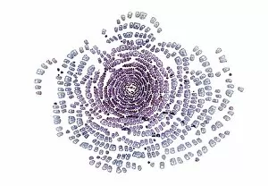
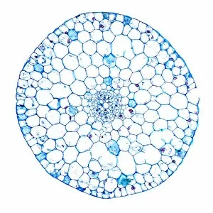
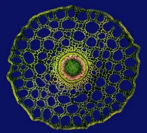
Cuticle Collection (#2)

"Exploring the Intricate World of Cuticles: From Water Lily Leaves to Giant Panda Hair" Delicate and ethereal

Background imageCuticle Collection: Hair, SEM

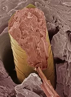
Hair, SEM
Hair fracture. Coloured scanning electron micrograph (SEM) of a fractured hair (yellow and pink). The surrounding layer of facial skin (brown) has a typically scale-like surface

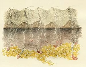
Background imageCuticle Collection: Palm

Palm. Magnified section of palm, historical artwork. The external ridged surface is the cuticle, with the thin layer of skin below this known as the rete mucosum, or epidermis

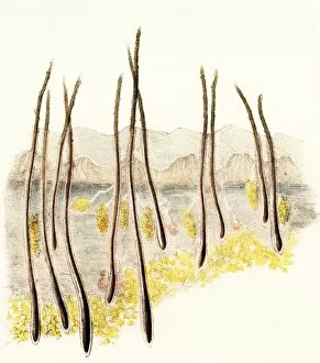
Background imageCuticle Collection: Magnified section of scalp

Magnified section of scalp, historical artwork. The external surface is the cuticle, with the layer of skin below this known as the rete mucosum, or epidermis

Background imageCuticle Collection: SEM of hair on scalp

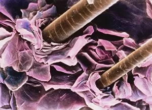
SEM of hair on scalp
Hair. Coloured scanning electron micrograph of hairs protruding from the surface of the scalp. Numerous desquamating cells (pink) are concentrically arranged around the base of the hair shaft

Background imageCuticle Collection: Coloured SEM of laser writing on a human hair

Coloured SEM of laser writing on a human hair
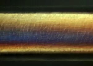
Laser script on human hair. Coloured scanning electron micrograph (SEM) of writing on a human hair performed by a medical laser

Background imageCuticle Collection: Nasturtium stem, SEM

Nasturtium stem, SEM
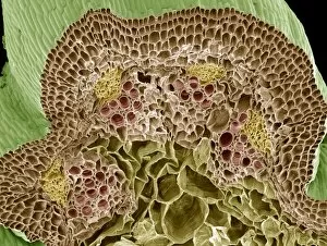
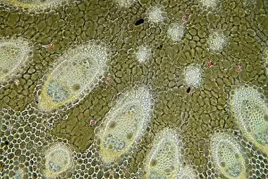
Nasturtium stem. Coloured scanning electron micrograph (SEM) of a freeze-fractured Nasturtium (Tropaeolum sp.) stem, showing numerous vascular bundles (such as at upper centre)

Background imageCuticle Collection: Water lily stem, SEM

Water lily stem, SEM
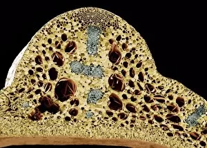
Water lily stem. Coloured scanning electron micrograph (SEM) of a freeze-fractured water lily stem showing numerous vascular bundles (grey) and large intercellular air spaces (holes)

Background imageCuticle Collection: Cashmere wool, SEM

Cashmere wool, SEM
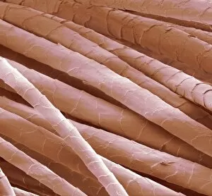
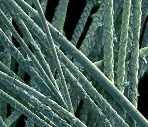
Cashmere wool. Coloured scanning electron micrograph (SEM) of hair fibres from the cashmere goat. The cuticle (outer layer) of the hair comprises overlapping scales

Background imageCuticle Collection: Common rush stem, light micrograph

Common rush stem, light micrograph
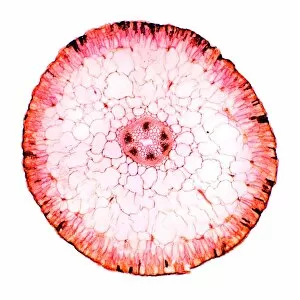
Common rush stem. Light micrograph of a section through the stem of a common rush (Juncus conglomeratus) plant, showing stellate cells

Background imageCuticle Collection: Sharp rush stem, light micrograph

Sharp rush stem, light micrograph
Sharp rush stem. Light micrograph of a section through the stem of a sharp rush (Juncus acutus) plant. This arid-adapted plant (xerophyte) has scattered vascular bundles

Background imageCuticle Collection: Ammophila arenaria leaf, light micrograph

Ammophila arenaria leaf, light micrograph
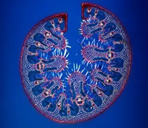
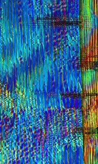
Ammophila arenaria leaf. Polarised light micrograph of a section through a marram grass (Ammophila arenaria) leaf, showing the characteristics that help reduce water loss

Background imageCuticle Collection: Hair shaft, light micrograph

Hair shaft, light micrograph
Hair shaft, differential interference contrast light micrograph. The outer layer of a hair shaft (the cuticle) has overlapping scales of keratin

Background imageCuticle Collection: Mint leaf surface, SEM

Mint leaf surface, SEM
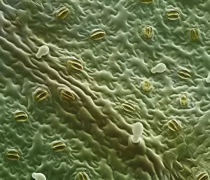
Mint leaf surface. Coloured scanning electron micrograph (SEM) of the surface of a mint (Menta sp.) leaf. The white structures are oil glands

Background imageCuticle Collection: Human hair, SEM

Human hair, SEM
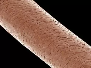
Human hair, coloured scanning electron micrograph (SEM). Hairs are made up of dead tissue. The outside of the hair, the cuticle

Background imageCuticle Collection: Oleander leaf, light micrograph

Oleander leaf, light micrograph
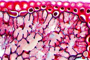
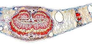
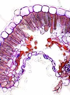
Oleander leaf. Light micrograph of a section through the leaf of an oleander (Nerium oleander) tree, showing its sunken stomata (gaps, centre left and right)

Background imageCuticle Collection: Pine tree needle, light micrograph

Pine tree needle, light micrograph
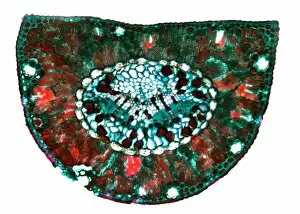
Pine tree needle. Polarised light micrograph of a cross-section through a needle from a Pinus pine tree. This leaf is needle-like in order to reduce water loss (transpiration)

Background imageCuticle Collection: Hair shaft anatomy, artwork

Hair shaft anatomy, artwork
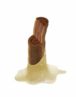
Hair shaft anatomy. Cutaway artwork showing the internal structure and anatomy of a human hair. Hair is made of a fibrous protein called keratin

Background imageCuticle Collection: Insect anatomy, artwork

Insect anatomy, artwork
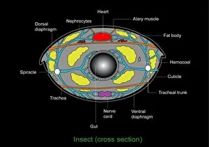
Insect anatomy. Computer artwork of a cross-section through the body of a typical Insect showing the main structures and organs. For the labelled diagram see image: C008/8724

Background imageCuticle Collection: Newly-emerged dragonflies

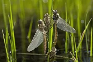
Newly-emerged dragonflies. Adult dragonflies (order Odonata) on plant stems after emerging from the nypmh stage. Their discarded nymphal cases (exuviae) can be seen below and between them

Background imageCuticle Collection: Kidney bean stem, light micrograph

Kidney bean stem, light micrograph
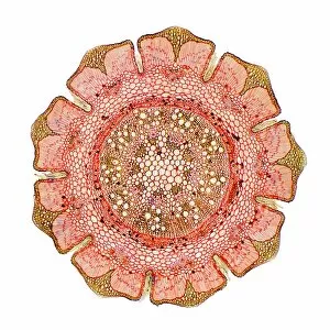
Kidney bean stem. Light micrograph of a section through the stem of a kidney bean (Phaseolus vulgaris) plant. The outer layer is the cuticle (brown), with a cortex of parenchyma (yellow) beneath it

Background imageCuticle Collection: Pine needle, light micrograph

Pine needle, light micrograph
Pine needle. Light micrograph of a transverse section through a leaf (needle) of a pine tree (Pinus sp.). The leaves are needle-like so they present a large surface area for photosynthesis but

Background imageCuticle Collection: Young pine tree stem, light micrograph

Young pine tree stem, light micrograph
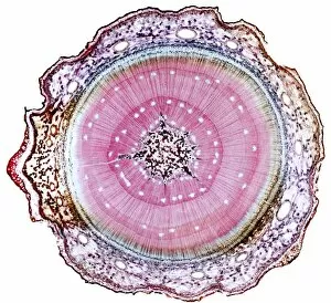
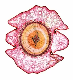
Young pine tree stem. Light micrograph of a transverse section through a two and a half-year-old stem of a pine tree (Pinus sp.)

Background imageCuticle Collection: Dyers greenweed stem, light micrograph

Dyers greenweed stem, light micrograph
Dyers greenweed stem. Polarised light micrograph of a transverse section through the stem of a dyers greenweed (Genista tinctoria) plant

Background imageCuticle Collection: Marsh samphire stem, light micrograph

Marsh samphire stem, light micrograph
Marsh samphire stem. Light micrograph of a transverse section through the stem of a marsh samphire, (Salicornia europaea) plant. This is a succulent, xerophytic, halophyte plant

Background imageCuticle Collection: Pine wood structure, light micrograph

Pine wood structure, light micrograph
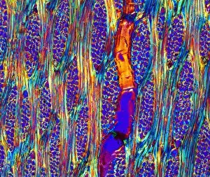
Pine wood structure. Polarised light micrograph of a longitudinal radial section through the stem wood (xylem) of the deal pine tree (Pinus mitis)

Background imageCuticle Collection: Eucalyptus leaf, light micrograph

Eucalyptus leaf, light micrograph
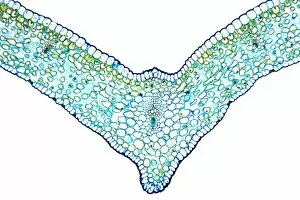
Eucalyptus leaf. Light micrograph of a cross-section through the midrib of a eucalyptus (Eucalyptus globulus) leaf. This xerophytic (arid-adapted)

Background imageCuticle Collection: Cedar tree stem, light micrograph

Cedar tree stem, light micrograph
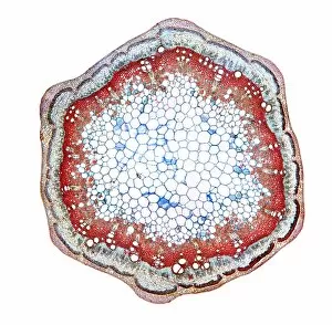
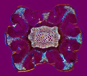
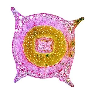
Cedar tree stem. Light micrograph of a transverse section through a stem of a cedar tree (Thujopsis dolobrata). The four ridges on the outer surface are microphyllous leaves

Background imageCuticle Collection: Sunflower stem, light micrograph

Sunflower stem, light micrograph
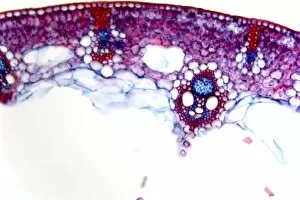
Sunflower stem. Light micrograph of a transverse section through the stem of a sunflower (Helianthus annuus) plant, showing a vascular bundle

Background imageCuticle Collection: Eucalyptus stem, light micrograph

Eucalyptus stem, light micrograph
Eucalyptus stem. Light micrograph of a transverse section through a one-year-old stem of a Eucalyptus (Eucalyptus globulus) plant

Background imageCuticle Collection: Mahogany wood structure, light micrograph

Mahogany wood structure, light micrograph
Mahogany wood structure. Polarised light micrograph of a longitudinal tangential section through a woody stem (xylem) of a mahogany (Swietenia sp.) tree

Background imageCuticle Collection: Beech leaves, light micrograph

Beech leaves, light micrograph
Beech leaves. Light micrograph of a transverse section through two beech leaves (Fagus sylvatica). The shapes of the two leaves are different because the bottom leaf is constantly exposed to bright
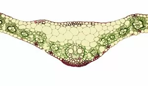
Background imageCuticle Collection: Ginger leaf, light micrograph

Ginger leaf, light micrograph
Ginger leaf. Light micrograph of a transverse section through the midrib of a ginger (Zingiber officinale) leaf. The lower and upper epidermis (blue)

Background imageCuticle Collection: Young yew tree stem, light micrograph

Young yew tree stem, light micrograph
Young yew tree stem. Light micrograph of a transverse section through a one-year-old stem of a yew tree (Taxus baccata). The epidermis (outer layer)

Background imageCuticle Collection: Cat hair, SEM

Cat hair, SEM
Cat hair, coloured scanning electron micrograph (SEM). The outside of the hair, the cuticle, is covered in overlapping scales of dead cells containing the protein keratin

Background imageCuticle Collection: Human hairs, SEM

Human hairs, SEM
Human hairs. Coloured scanning electron micrograph (SEM) of three human hairs next to each other. The outside of the hair, the cuticle
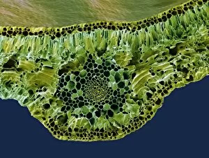
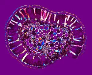
Background imageCuticle Collection: Leaf midrib, light micrograph

Leaf midrib, light micrograph
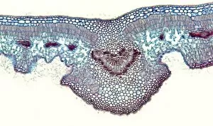
Leaf midrib. Light micrograph (LM) of a section through the midrib of a leaf from a monocotyledon plant. The midrib (midvein) is the continuation of a leafs stem along the centre of the leaf

Background imageCuticle Collection: Leaf midrib, SEM

Leaf midrib, SEM
Leaf midrib. Coloured scanning electron micrograph (SEM) of a section through the midrib of a leaf from the Common Box (Buxus sempervirens)

Background imageCuticle Collection: Pincushion hakea leaf, light micrograph

Pincushion hakea leaf, light micrograph
Pincushion hakea leaf. Polarised light micrograph of a transverse section through the leaf of a pincushion hakea (Hakea laurina) plant. This is a drought plant (xerophyte)

Background imageCuticle Collection: Heather leaf stomata, light micrograph

Heather leaf stomata, light micrograph
Heather leaf stomata. Light micrograph of a transverse section through a stomata (centre, circular) in the leaf of a heather (Erica sp.) plant. Heather is a drought plant (xerophyte)

Background imageCuticle Collection: Heather leaf, light micrograph

Heather leaf, light micrograph
Heather leaf. Light micrograph of a transverse section through the leaf of a heather (Erica sp.) plant. Heather is a drought plant (xerophyte)

Background imageCuticle Collection: She-oak stem, light micrograph

She-oak stem, light micrograph
She-oak stem. Light micrograph of a transverse section through the stem of the Australian she-oak (Casuarina equisetifolia). The she-oak is a drought plant (xerophyte)

Background imageCuticle Collection: Water lily leaf stalk, light micrograph

Water lily leaf stalk, light micrograph
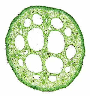
Water lily leaf stalk. Light micrograph of a transverse section through the leaf stalk (petiole) of a water lily (Nymphaea sp.). All aquatic plants (hydrophytes) have a similar structure

Background imageCuticle Collection: Pondweed stem, light micrograph

Pondweed stem, light micrograph
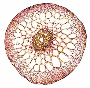
Pondweed stem. Light micrograph of a transverse section through the stem of a pondweed (Potamogeton sp.) plant. All aquatic plants (hydrophytes) have a similar stem structure

Background imageCuticle Collection: Water milfoil stem, light micrograph

Water milfoil stem, light micrograph
Water milfoil stem. Light micrograph of a transverse section through the stem of the aquatic whorled water milfoil (Myriophyllum verticillatum) plant

Background imageCuticle Collection: Water milfoil shoot-tip, light micrograph

Water milfoil shoot-tip, light micrograph
Water milfoil shoot tip. Light micrograph of a transverse section through the shoot-tip of the aquatic whorled water milfoil (Myriophyllum verticillatum) plant

Background imageCuticle Collection: Waterweed stem, light micrograph

Waterweed stem, light micrograph
Waterweed stem. Polarised light micrograph of a transverse section through a stem of the aquatic western waterweed (Elodea nuttallii) plant

Background imageCuticle Collection: Mistletoe stem, light micrograph

Mistletoe stem, light micrograph
Mistletoe stem. Polarised light micrograph of a transverse section through the stem of a mistletoe (Viscum album) plant. The epidermis (outer layer)

Background imageCuticle Collection: Mares tail stem, light micrograph

Mares tail stem, light micrograph
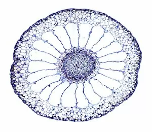
Mares tail stem. Polarised light micrograph of a transverse section through a stem of the aquatic mares tail (Hippuris vulgaris) plant. All aquatic plants (hydrophytes) have a similar stem structure


For sale as Licensed Images

Choose your image, Select your licence and Download the media

"Exploring the Intricate World of Cuticles: From Water Lily Leaves to Giant Panda Hair" Delicate and ethereal, the water lily leaf showcases its intricate cuticle structure under a light micrograph. Unveiling the evolution of beauty culture, a vintage black and white photo captures the electric manicure machine revolutionizing nail care. Nature's artistry at play: glistening water droplets delicately rest on a leaf's cuticle, creating an enchanting sight. Peering into the microscopic world, a biomedical illustration reveals the complex anatomy of a roundworm's cuticle – nature's armor for survival. Human hair takes center stage as scanning electron microscopy (SEM) unravels its fascinating structural details with astonishing clarity. Not just limited to humans, SEM delves into dog hair too, unraveling its unique composition and texture in mesmerizing detail. A captivating SEM image showcases giant panda hair in all its glory – each strand revealing nature's mastery in providing insulation and protection. Gastrotrichs take their turn under SEM scrutiny, showcasing their intriguing cuticular features that aid them in aquatic habitats. Another glimpse into human hair through SEM highlights its diverse textures and patterns – truly reflecting individuality at its finest. In this captivating journey through various specimens captured by cutting-edge technology like SEM or traditional photography methods like light micrographs, we discover how different organisms possess unique yet equally fascinating cuticles - from delicate leaves to human hairs or even gastrotrichs thriving underwater. The intricacies hidden within these seemingly ordinary structures remind us of nature’s remarkable ability to adapt and protect living beings across species boundaries.